 Movie
Movie Controller

[English] 日本語
 Yorodumi
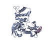
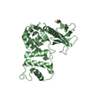
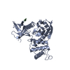
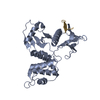
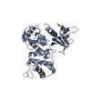
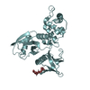
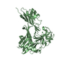
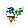
- PDB-2zpy: Crystal structure of the mouse radxin FERM domain complexed with ... -
+ Open data

- Basic information
| Entry | Database: PDB / ID: 2zpy | ||||||
|---|---|---|---|---|---|---|---|
| Title | Crystal structure of the mouse radxin FERM domain complexed with the mouse CD44 cytoplasmic peptide | ||||||
 Components Components |
| ||||||
 Keywords Keywords | CELL ADHESION / FERM domain / CD44 / Actin capping / Actin-binding / Cell membrane / Cytoplasm / Cytoskeleton / Membrane / Phosphoprotein / Structural protein / Alternative splicing / Glycoprotein / Proteoglycan / Pyrrolidone carboxylic acid / Receptor / Sulfation / Transmembrane | ||||||
| Function / homology |  Function and homology information Function and homology informationregulation of adherens junction organization / stereocilium base / regulation of organelle assembly / microvillus assembly / establishment of protein localization to plasma membrane / Hyaluronan metabolism / Hyaluronan degradation / positive regulation of monocyte aggregation / positive regulation of early endosome to late endosome transport / macrophage fusion ...regulation of adherens junction organization / stereocilium base / regulation of organelle assembly / microvillus assembly / establishment of protein localization to plasma membrane / Hyaluronan metabolism / Hyaluronan degradation / positive regulation of monocyte aggregation / positive regulation of early endosome to late endosome transport / macrophage fusion / hyaluronic acid binding / macrophage migration inhibitory factor receptor complex / regulation of Rap protein signal transduction / Degradation of the extracellular matrix / Integrin cell surface interactions / regulation of lamellipodium morphogenesis / postsynapse organization / Cell surface interactions at the vascular wall / Recycling pathway of L1 / monocyte aggregation / wound healing involved in inflammatory response / positive regulation of protein localization to early endosome / hyaluronan catabolic process / : / regulation of postsynaptic neurotransmitter receptor diffusion trapping / branching involved in prostate gland morphogenesis / cell tip / positive regulation of adaptive immune response / type II transforming growth factor beta receptor binding / negative regulation of CD4-positive, alpha-beta T cell proliferation / negative regulation of mature B cell apoptotic process / positive regulation of neutrophil apoptotic process / barbed-end actin filament capping / stereocilium / regulation of modification of postsynaptic structure / cellular response to thyroid hormone stimulus / channel regulator activity / positive regulation of heterotypic cell-cell adhesion / establishment of endothelial barrier / wound healing, spreading of cells / apical protein localization / epidermal growth factor receptor binding / cargo receptor activity / branching involved in ureteric bud morphogenesis / negative regulation of regulatory T cell differentiation / negative regulation of intrinsic apoptotic signaling pathway in response to DNA damage by p53 class mediator / cortical actin cytoskeleton / negative regulation of DNA damage response, signal transduction by p53 class mediator / protein kinase A binding / microvillus / cleavage furrow / lamellipodium membrane / positive regulation of G1/S transition of mitotic cell cycle / cellular response to platelet-derived growth factor stimulus / ruffle / cell projection / Neutrophil degranulation / cellular response to fibroblast growth factor stimulus / cell adhesion molecule binding / T-tubule / receptor-mediated endocytosis / protein localization to plasma membrane / cell periphery / T cell activation / filopodium / adherens junction / establishment of protein localization / phosphoprotein binding / regulation of cell growth / negative regulation of inflammatory response / Wnt signaling pathway / neuron projection development / cytokine-mediated signaling pathway / apical part of cell / positive regulation of protein catabolic process / transmembrane signaling receptor activity / myelin sheath / regulation of cell shape / cell migration / lamellipodium / ATPase binding / presynapse / actin binding / midbody / basolateral plasma membrane / positive regulation of ERK1 and ERK2 cascade / cell adhesion / postsynapse / apical plasma membrane / membrane raft / inflammatory response / external side of plasma membrane / protein domain specific binding / focal adhesion / positive regulation of gene expression / protein kinase binding / negative regulation of apoptotic process / glutamatergic synapse / Golgi apparatus / cell surface Similarity search - Function | ||||||
| Biological species |  | ||||||
| Method |  X-RAY DIFFRACTION / SYNCHROTRON / MOLECULAR REPLACEMENT / Resolution: 2.1 Å X-RAY DIFFRACTION / SYNCHROTRON / MOLECULAR REPLACEMENT / Resolution: 2.1 Å | ||||||
 Authors Authors | Mori, T. / Kitano, K. / Terawaki, S. / Maesaki, R. / Fukami, Y. / Hakoshima, T. | ||||||
 Citation Citation | Journal: J.Biol.Chem. / Year: 2008 Title: Structural basis for CD44 recognition by ERM proteins Authors: Mori, T. / Kitano, K. / Terawaki, S. / Maesaki, R. / Fukami, Y. / Hakoshima, T. | ||||||
| History |
|
- Structure visualization
| Structure viewer | Molecule: MolmilJmol/JSmol |
|---|
- Downloads & links
-Download
| PDBx/mmCIF format | 2zpy.cif.gz | 80.1 KB | Display | PDBx/mmCIF format |
|---|---|---|---|---|
| PDB format | pdb2zpy.ent.gz | 58.9 KB | Display | PDB format |
| PDBx/mmJSON format | 2zpy.json.gz | Tree view | PDBx/mmJSON format | |
| Others |  Other downloads Other downloads |
-Validation report
| Arichive directory | https://data.pdbj.org/pub/pdb/validation_reports/zp/2zpyftp://data.pdbj.org/pub/pdb/validation_reports/zp/2zpy | HTTPS FTP |
|---|
-Related structure data
| Related structure data |  1gc7S S: Starting model for refinement |
|---|---|
| Similar structure data |


-Links
 PDBj
PDBj


- Assembly
| Deposited unit | 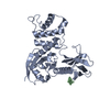
| ||||||||
|---|---|---|---|---|---|---|---|---|---|
| 1 |
| ||||||||
| Unit cell |
|
-Components
| #1: Protein | Mass: 36934.598 Da / Num. of mol.: 1 / Fragment: FERM domain (residues 1-310) Source method: isolated from a genetically manipulated source Source: (gene. exp.)  |
|---|---|
| #2: Protein/peptide | Mass: 2177.579 Da / Num. of mol.: 1 / Fragment: residues 293-312 / Source method: obtained synthetically / Details: peptide synthesis / References: UniProt: P15379 |
| #3: Water | ChemComp-HOH /  Mass: 18.015 Da / Num. of mol.: 171 / Source method: isolated from a natural source / Formula: H2O Mass: 18.015 Da / Num. of mol.: 171 / Source method: isolated from a natural source / Formula: H2O |
-Experimental details
-Experiment
| Experiment | Method: X-RAY DIFFRACTION / Number of used crystals: 1 |
|---|
- Sample preparation
| Crystal | Density Matthews: 2.29 Å3/Da / Density % sol: 46.21 % |
|---|---|
| Crystal grow | Temperature: 277 K / Method: vapor diffusion / pH: 8.6 Details: 0.1M Tris, 0.2M Potassium thiocyanate, 15% PEG 3350 , pH 8.6, VAPOR DIFFUSION, temperature 277K |
-Data collection
| Diffraction | Mean temperature: 100 K |
|---|---|
| Diffraction source | Source: SYNCHROTRON / Site: SPring-8  / Beamline: BL38B1 / Wavelength: 1 Å / Beamline: BL38B1 / Wavelength: 1 Å |
| Detector | Type: MAR CCD 165 mm / Detector: CCD / Date: Oct 17, 2006 |
| Radiation | Protocol: SINGLE WAVELENGTH / Monochromatic (M) / Laue (L): M / Scattering type: x-ray |
| Radiation wavelength | Wavelength: 1 Å / Relative weight: 1 |
| Reflection | Resolution: 2.1→20 Å / Num. obs: 21492 / % possible obs: 100 % |
| Reflection shell | Resolution: 2.1→2.17 Å / % possible all: 100 |
- Processing
| Software |
| ||||||||||||||||
|---|---|---|---|---|---|---|---|---|---|---|---|---|---|---|---|---|---|
| Refinement | Method to determine structure: MOLECULAR REPLACEMENT Starting model: 1GC7 Resolution: 2.1→20 Å
| ||||||||||||||||
| Refinement step | Cycle: LAST / Resolution: 2.1→20 Å
|
