 Movie
Movie Controller


+ Open data

- Basic information
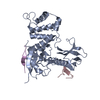
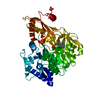
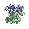
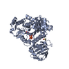
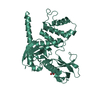
| Entry | Database: PDB / ID: 1isn | ||||||
|---|---|---|---|---|---|---|---|
| Title | Crystal structure of merlin FERM domain | ||||||
 Components Components | merlin | ||||||
 Keywords Keywords | CELL ADHESION / FERM domain | ||||||
| Function / homology |  Function and homology information Function and homology informationregulation of hippo signaling / RHO GTPases activate PAKs / regulation of organelle assembly / positive regulation of early endosome to late endosome transport / Schwann cell proliferation / regulation of gliogenesis / regulation of stem cell proliferation / ectoderm development / lens fiber cell differentiation / Regulation of actin dynamics for phagocytic cup formation ...regulation of hippo signaling / RHO GTPases activate PAKs / regulation of organelle assembly / positive regulation of early endosome to late endosome transport / Schwann cell proliferation / regulation of gliogenesis / regulation of stem cell proliferation / ectoderm development / lens fiber cell differentiation / Regulation of actin dynamics for phagocytic cup formation / positive regulation of protein localization to early endosome / regulation of neural precursor cell proliferation / cell-cell junction organization / negative regulation of receptor signaling pathway via JAK-STAT / regulation of protein localization to nucleus / negative regulation of cell-cell adhesion / odontogenesis of dentin-containing tooth / cortical actin cytoskeleton / cleavage furrow / mesoderm formation / regulation of neurogenesis / negative regulation of MAPK cascade / ruffle / positive regulation of stress fiber assembly / hippocampus development / filopodium / adherens junction / positive regulation of cell differentiation / regulation of protein stability / brain development / negative regulation of cell growth / beta-catenin binding / integrin binding / apical part of cell / regulation of cell population proliferation / regulation of cell shape / lamellipodium / actin cytoskeleton organization / actin binding / cell body / regulation of apoptotic process / cytoskeleton / early endosome / regulation of cell cycle / neuron projection / negative regulation of cell population proliferation / protein domain specific binding / nucleolus / perinuclear region of cytoplasm / protein-containing complex / nucleus / plasma membrane / cytosol / cytoplasm Similarity search - Function | ||||||
| Biological species |  | ||||||
| Method |  X-RAY DIFFRACTION / SYNCHROTRON / MOLECULAR REPLACEMENT / Resolution: 2.9 Å X-RAY DIFFRACTION / SYNCHROTRON / MOLECULAR REPLACEMENT / Resolution: 2.9 Å | ||||||
 Authors Authors | Shimizu, T. / Seto, A. / Maita, N. / Hamada, K. / Tsukita, S. / Tsukita, S. / Hakoshima, T. | ||||||
 Citation Citation | Journal: J.Biol.Chem. / Year: 2002 Title: Structural basis for neurofibromatosis type 2. Crystal structure of the merlin FERM domain. Authors: Shimizu, T. / Seto, A. / Maita, N. / Hamada, K. / Tsukita, S. / Tsukita, S. / Hakoshima, T. | ||||||
| History |
|
- Structure visualization
| Structure viewer | Molecule: MolmilJmol/JSmol |
|---|
- Downloads & links
-Download
| PDBx/mmCIF format | 1isn.cif.gz | 77.2 KB | Display | PDBx/mmCIF format |
|---|---|---|---|---|
| PDB format | pdb1isn.ent.gz | 57.5 KB | Display | PDB format |
| PDBx/mmJSON format | 1isn.json.gz | Tree view | PDBx/mmJSON format | |
| Others |  Other downloads Other downloads |
-Validation report
| Arichive directory | https://data.pdbj.org/pub/pdb/validation_reports/is/1isnftp://data.pdbj.org/pub/pdb/validation_reports/is/1isn | HTTPS FTP |
|---|
-Related structure data
| Related structure data |  1gc6S S: Starting model for refinement |
|---|---|
| Similar structure data |


-Links
 PDBj
PDBj


- Assembly
| Deposited unit | 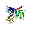
| ||||||||
|---|---|---|---|---|---|---|---|---|---|
| 1 |
| ||||||||
| Unit cell |
|
-Components
| #1: Protein | Mass: 38367.320 Da / Num. of mol.: 1 / Fragment: FERM domain Source method: isolated from a genetically manipulated source Source: (gene. exp.)  |
|---|
-Experimental details
-Experiment
| Experiment | Method: X-RAY DIFFRACTION / Number of used crystals: 1 |
|---|
- Sample preparation
| Crystal | Density Matthews: 2.65 Å3/Da / Density % sol: 53.6 % | ||||||||||||||||||||||||||||||||||||||||||||||||||||||||||||||||||||||
|---|---|---|---|---|---|---|---|---|---|---|---|---|---|---|---|---|---|---|---|---|---|---|---|---|---|---|---|---|---|---|---|---|---|---|---|---|---|---|---|---|---|---|---|---|---|---|---|---|---|---|---|---|---|---|---|---|---|---|---|---|---|---|---|---|---|---|---|---|---|---|---|
| Crystal grow | Temperature: 277 K / Method: vapor diffusion, hanging drop / pH: 7 Details: PEG6000, LiCl, pH 7.0, VAPOR DIFFUSION, HANGING DROP, temperature 277K | ||||||||||||||||||||||||||||||||||||||||||||||||||||||||||||||||||||||
| Crystal grow | *PLUS Temperature: 4 ℃ / pH: 6.8 | ||||||||||||||||||||||||||||||||||||||||||||||||||||||||||||||||||||||
| Components of the solutions | *PLUS
|
-Data collection
| Diffraction | Mean temperature: 93 K |
|---|---|
| Diffraction source | Source: SYNCHROTRON / Site: SPring-8  / Beamline: BL41XU / Wavelength: 0.9198 Å / Beamline: BL41XU / Wavelength: 0.9198 Å |
| Detector | Type: MARRESEARCH / Detector: CCD / Date: Nov 10, 2000 |
| Radiation | Monochromator: double-crystal / Protocol: SINGLE WAVELENGTH / Monochromatic (M) / Laue (L): M / Scattering type: x-ray |
| Radiation wavelength | Wavelength: 0.9198 Å / Relative weight: 1 |
| Reflection | Resolution: 2.9→30 Å / Num. all: 8825 / Num. obs: 8807 / % possible obs: 90.5 % / Observed criterion σ(F): 0 / Observed criterion σ(I): 0 / Redundancy: 5.5 % / Biso Wilson estimate: 50.8 Å2 / Rmerge(I) obs: 0.069 / Net I/σ(I): 8.4 |
| Reflection shell | Resolution: 2.9→3.03 Å / Rmerge(I) obs: 0.316 / Mean I/σ(I) obs: 2.4 / % possible all: 75.8 |
| Reflection | *PLUS Lowest resolution: 30 Å / Num. measured all: 32050 / Rmerge(I) obs: 0.069 |
| Reflection shell | *PLUS % possible obs: 75.8 % / Rmerge(I) obs: 0.316 |
- Processing
| Software |
| |||||||||||||||||||||||||
|---|---|---|---|---|---|---|---|---|---|---|---|---|---|---|---|---|---|---|---|---|---|---|---|---|---|---|
| Refinement | Method to determine structure: MOLECULAR REPLACEMENT Starting model: PDB ENTRY 1GC6 Resolution: 2.9→20 Å / Cross valid method: THROUGHOUT / σ(F): 2 / Stereochemistry target values: Engh & Huber
| |||||||||||||||||||||||||
| Refine analyze |
| |||||||||||||||||||||||||
| Refinement step | Cycle: LAST / Resolution: 2.9→20 Å
| |||||||||||||||||||||||||
| Refine LS restraints |
| |||||||||||||||||||||||||
| LS refinement shell | Resolution: 2.9→3.03 Å
| |||||||||||||||||||||||||
| Refinement | *PLUS Highest resolution: 2.9 Å / Lowest resolution: 20 Å / σ(F): 2 / % reflection Rfree: 10 % / Rfactor all: 0.237 / Rfactor obs: 0.229 / Rfactor Rfree: 0.284 / Rfactor Rwork: 0.229 | |||||||||||||||||||||||||
| Solvent computation | *PLUS | |||||||||||||||||||||||||
| Displacement parameters | *PLUS | |||||||||||||||||||||||||
| Refine LS restraints | *PLUS
| |||||||||||||||||||||||||
| LS refinement shell | *PLUS Highest resolution: 2.9 Å / Rfactor Rfree: 0.4065 / Rfactor Rwork: 0.3728 / Rfactor obs: 0.373 |
