 Movie
Movie Controller

[English] 日本語
 Yorodumi
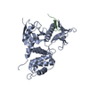
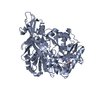
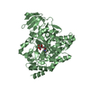
- PDB-1j19: Crystal structure of the radxin FERM domain complexed with the IC... -
+ Open data

- Basic information
| Entry | Database: PDB / ID: 1j19 | ||||||
|---|---|---|---|---|---|---|---|
| Title | Crystal structure of the radxin FERM domain complexed with the ICAM-2 cytoplasmic peptide | ||||||
 Components Components |
| ||||||
 Keywords Keywords | CELL ADHESION / protein-peptide complex | ||||||
| Function / homology |  Function and homology information Function and homology informationregulation of adherens junction organization / stereocilium base / regulation of organelle assembly / dendritic cell migration / microvillus assembly / establishment of protein localization to plasma membrane / regulation of Rap protein signal transduction / positive regulation of early endosome to late endosome transport / uropod / Integrin cell surface interactions ...regulation of adherens junction organization / stereocilium base / regulation of organelle assembly / dendritic cell migration / microvillus assembly / establishment of protein localization to plasma membrane / regulation of Rap protein signal transduction / positive regulation of early endosome to late endosome transport / uropod / Integrin cell surface interactions / Recycling pathway of L1 / positive regulation of protein localization to early endosome / CD209 (DC-SIGN) signaling / regulation of postsynaptic neurotransmitter receptor diffusion trapping / cell tip / barbed-end actin filament capping / stereocilium / cellular response to thyroid hormone stimulus / apical protein localization / establishment of endothelial barrier / Immunoregulatory interactions between a Lymphoid and a non-Lymphoid cell / cell adhesion mediated by integrin / leukocyte cell-cell adhesion / cortical actin cytoskeleton / protein kinase A binding / microvillus / cleavage furrow / positive regulation of G1/S transition of mitotic cell cycle / ruffle / cell adhesion molecule binding / T-tubule / protein localization to plasma membrane / cell periphery / filopodium / adherens junction / establishment of protein localization / integrin binding / apical part of cell / positive regulation of protein catabolic process / myelin sheath / regulation of cell shape / lamellipodium / ATPase binding / actin binding / midbody / cell adhesion / apical plasma membrane / receptor ligand activity / protein domain specific binding / focal adhesion / positive regulation of gene expression / plasma membrane / cytosol Similarity search - Function | ||||||
| Biological species |  | ||||||
| Method |  X-RAY DIFFRACTION / SYNCHROTRON / MOLECULAR REPLACEMENT / Resolution: 2.4 Å X-RAY DIFFRACTION / SYNCHROTRON / MOLECULAR REPLACEMENT / Resolution: 2.4 Å | ||||||
 Authors Authors | Hamada, K. / Shimizu, T. / Yonemura, S. / Tsukita, S. / Tsukita, S. / Hakoshima, T. | ||||||
 Citation Citation | Journal: EMBO J. / Year: 2003 Title: Structural basis of adhesion-molecule recognition by ERM proteins revealed by the crystal structure of the radixin-ICAM-2 complex Authors: Hamada, K. / Shimizu, T. / Yonemura, S. / Tsukita, S. / Tsukita, S. / Hakoshima, T. #1: Journal: Acta Crystallogr.,Sect.D / Year: 2001Title: Crystallographic characterization of the radixin FERM domain bound to the cytoplasmic tail of the adhesion protein ICAM-2 Authors: Hamada, K. / Shimizu, T. / Matsui, T. / Tsukita, S. / Tsukita, S. / Hakoshima, T. | ||||||
| History |
|
- Structure visualization
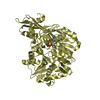
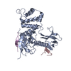
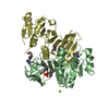
| Structure viewer | Molecule: MolmilJmol/JSmol |
|---|
- Downloads & links
-Download
| PDBx/mmCIF format | 1j19.cif.gz | 84.9 KB | Display | PDBx/mmCIF format |
|---|---|---|---|---|
| PDB format | pdb1j19.ent.gz | 63.8 KB | Display | PDB format |
| PDBx/mmJSON format | 1j19.json.gz | Tree view | PDBx/mmJSON format | |
| Others |  Other downloads Other downloads |
-Validation report
| Arichive directory | https://data.pdbj.org/pub/pdb/validation_reports/j1/1j19ftp://data.pdbj.org/pub/pdb/validation_reports/j1/1j19 | HTTPS FTP |
|---|
-Related structure data
| Related structure data |  1gc7S S: Starting model for refinement |
|---|---|
| Similar structure data |


-Links
 PDBj
PDBj


- Assembly
| Deposited unit | 
| ||||||||
|---|---|---|---|---|---|---|---|---|---|
| 1 |
| ||||||||
| Unit cell |
|
-Components
| #1: Protein | Mass: 37368.008 Da / Num. of mol.: 1 / Fragment: N-terminal domain, FERM domain Source method: isolated from a genetically manipulated source Source: (gene. exp.)  |
|---|---|
| #2: Protein/peptide | Mass: 1937.281 Da / Num. of mol.: 1 Source method: isolated from a genetically manipulated source Source: (gene. exp.) |
| #3: Water | ChemComp-HOH /  Mass: 18.015 Da / Num. of mol.: 232 / Source method: isolated from a natural source / Formula: H2O Mass: 18.015 Da / Num. of mol.: 232 / Source method: isolated from a natural source / Formula: H2O |
-Experimental details
-Experiment
| Experiment | Method: X-RAY DIFFRACTION / Number of used crystals: 2 |
|---|
- Sample preparation
| Crystal | Density Matthews: 3.49 Å3/Da / Density % sol: 64.46 % | ||||||||||||||||||||||||||||||||||||||||||
|---|---|---|---|---|---|---|---|---|---|---|---|---|---|---|---|---|---|---|---|---|---|---|---|---|---|---|---|---|---|---|---|---|---|---|---|---|---|---|---|---|---|---|---|
| Crystal grow | Temperature: 277 K / Method: vapor diffusion, hanging drop / pH: 6 Details: PEG6000, MES, NaCl, DTT, pH 6.0, VAPOR DIFFUSION, HANGING DROP, temperature 277K | ||||||||||||||||||||||||||||||||||||||||||
| Crystal grow | *PLUS Temperature: 4 ℃ / Method: unknown | ||||||||||||||||||||||||||||||||||||||||||
| Components of the solutions | *PLUS
|
-Data collection
| Diffraction | Mean temperature: 100 K |
|---|---|
| Diffraction source | Source: SYNCHROTRON / Site: SPring-8  / Beamline: BL41XU / Wavelength: 1 Å / Beamline: BL41XU / Wavelength: 1 Å |
| Detector | Type: MARRESEARCH / Detector: CCD |
| Radiation | Protocol: SINGLE WAVELENGTH / Monochromatic (M) / Laue (L): M / Scattering type: x-ray |
| Radiation wavelength | Wavelength: 1 Å / Relative weight: 1 |
| Reflection | Resolution: 2.4→44.7 Å / Num. all: 599708 / Num. obs: 23153 / % possible obs: 100 % / Biso Wilson estimate: 41.7 Å2 |
| Reflection shell | Resolution: 2.4→2.53 Å / Rmerge(I) obs: 0.137 / Mean I/σ(I) obs: 5.2 / % possible all: 100 |
| Reflection | *PLUS Highest resolution: 2.4 Å / % possible obs: 100 % / Num. measured all: 599708 / Rmerge(I) obs: 0.071 |
| Reflection shell | *PLUS % possible obs: 100 % |
- Processing
| Software |
| ||||||||||||||||||||||||||||||||||||||||
|---|---|---|---|---|---|---|---|---|---|---|---|---|---|---|---|---|---|---|---|---|---|---|---|---|---|---|---|---|---|---|---|---|---|---|---|---|---|---|---|---|---|
| Refinement | Method to determine structure: MOLECULAR REPLACEMENT Starting model: PDB ENTRY 1GC7 Resolution: 2.4→14.94 Å / Rfactor Rfree error: 0.005 / Data cutoff high absF: 1411875 / Data cutoff low absF: 0 / Isotropic thermal model: RESTRAINED / Cross valid method: THROUGHOUT / σ(F): 0 / Stereochemistry target values: Engh & Huber
| ||||||||||||||||||||||||||||||||||||||||
| Solvent computation | Solvent model: FLAT MODEL / Bsol: 65.0264 Å2 / ksol: 0.328436 e/Å3 | ||||||||||||||||||||||||||||||||||||||||
| Displacement parameters | Biso mean: 52.3 Å2
| ||||||||||||||||||||||||||||||||||||||||
| Refine analyze |
| ||||||||||||||||||||||||||||||||||||||||
| Refinement step | Cycle: LAST / Resolution: 2.4→14.94 Å
| ||||||||||||||||||||||||||||||||||||||||
| Refine LS restraints |
| ||||||||||||||||||||||||||||||||||||||||
| LS refinement shell | Resolution: 2.4→2.55 Å / Rfactor Rfree error: 0.019 / Total num. of bins used: 6
| ||||||||||||||||||||||||||||||||||||||||
| Xplor file |
| ||||||||||||||||||||||||||||||||||||||||
| Refinement | *PLUS Lowest resolution: 15 Å / % reflection Rfree: 10 % / Rfactor obs: 0.229 / Rfactor Rfree: 0.24 / Rfactor Rwork: 0.229 | ||||||||||||||||||||||||||||||||||||||||
| Solvent computation | *PLUS | ||||||||||||||||||||||||||||||||||||||||
| Displacement parameters | *PLUS | ||||||||||||||||||||||||||||||||||||||||
| Refine LS restraints | *PLUS
| ||||||||||||||||||||||||||||||||||||||||
| LS refinement shell | *PLUS Lowest resolution: 2.53 Å |
