[English] 日本語
Yorodumi
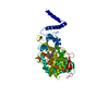
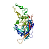
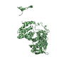
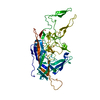
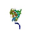
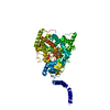
- PDB-1e5w: Structure of isolated FERM domain and first long helix of moesin -

+
Open data


ID or keywords:

Loading...

-
Basic information

Entry
Database: PDB / ID: 1e5w
TitleStructure of isolated FERM domain and first long helix of moesin
ComponentsMOESIN
KeywordsMEMBRANE PROTEIN / MOESIN / FERM / ERM
Function / homology
Function and homology information


regulation of lymphocyte migration / T cell aggregation / regulation of organelle assembly / T cell migration / positive regulation of early endosome to late endosome transport / membrane to membrane docking / uropod / immunological synapse formation / gland morphogenesis / positive regulation of protein localization to early endosome ...regulation of lymphocyte migration / T cell aggregation / regulation of organelle assembly / T cell migration / positive regulation of early endosome to late endosome transport / membrane to membrane docking / uropod / immunological synapse formation / gland morphogenesis / positive regulation of protein localization to early endosome / cellular response to testosterone stimulus / establishment of epithelial cell apical/basal polarity / establishment of endothelial barrier / Sensory processing of sound by outer hair cells of the cochlea / positive regulation of podosome assembly / Sensory processing of sound by inner hair cells of the cochlea / microvillus membrane / leukocyte cell-cell adhesion / regulation of cell size / leukocyte migration / pseudopodium / Recycling pathway of L1 / microvillus / T cell proliferation / cell adhesion molecule binding / Gene and protein expression by JAK-STAT signaling after Interleukin-12 stimulation / filopodium / cell periphery / adherens junction / structural constituent of cytoskeleton / positive regulation of protein catabolic process / double-stranded RNA binding / Signaling by ALK fusions and activated point mutants / apical part of cell / actin binding / regulation of cell shape / basolateral plasma membrane / vesicle / cytoskeleton / blood microparticle / apical plasma membrane / focal adhesion / signaling receptor binding / positive regulation of gene expression / protein kinase binding / perinuclear region of cytoplasm / enzyme binding / cell surface / extracellular space / extracellular exosome / nucleus / plasma membrane / cytoplasm / cytosol
Similarity search - Function
Single alpha-helices involved in coiled-coils or other helix-helix interfaces - #450 / Ezrin/radixin/moesin, alpha-helical domain / Ezrin/radixin/moesin, alpha-helical domain / Moesin tail domain superfamily / Ezrin/radixin/moesin / Ezrin/radixin/moesin, C-terminal / ERM family, FERM domain C-lobe / Ezrin/radixin/moesin family C terminal / Acyl-CoA Binding Protein - #10 / Ezrin/radixin/moesin-like ...Single alpha-helices involved in coiled-coils or other helix-helix interfaces - #450 / Ezrin/radixin/moesin, alpha-helical domain / Ezrin/radixin/moesin, alpha-helical domain / Moesin tail domain superfamily / Ezrin/radixin/moesin / Ezrin/radixin/moesin, C-terminal / ERM family, FERM domain C-lobe / Ezrin/radixin/moesin family C terminal / Acyl-CoA Binding Protein - #10 / Ezrin/radixin/moesin-like / Acyl-CoA Binding Protein / FERM, C-terminal PH-like domain / FERM C-terminal PH-like domain / FERM C-terminal PH-like domain / FERM, N-terminal / FERM N-terminal domain / FERM domain signature 1. / FERM conserved site / FERM domain signature 2. / FERM central domain / FERM/acyl-CoA-binding protein superfamily / Pleckstrin-homology domain (PH domain)/Phosphotyrosine-binding domain (PTB) / PH-domain like / FERM central domain / FERM superfamily, second domain / FERM domain / FERM domain profile. / Band 4.1 domain / Band 4.1 homologues / Phosphatidylinositol 3-kinase Catalytic Subunit; Chain A, domain 1 / Single alpha-helices involved in coiled-coils or other helix-helix interfaces / Ubiquitin-like (UB roll) / PH-like domain superfamily / Ubiquitin-like domain superfamily / Roll / Roll / Up-down Bundle / Mainly Beta / Mainly Alpha / Alpha Beta
Similarity search - Domain/homology
Biological speciesHOMO SAPIENS (human)
MethodX-RAY DIFFRACTION / SYNCHROTRON / MAD / Resolution: 2.7 Å
AuthorsEdwards, S.D. / Keep, N.H.
CitationJournal: Biochemistry / Year: 2001
Title: The 2.7 A Crystal Structure of the Activated Ferm Domain of Moesin: An Analysis of Structural Changes on Activation
Authors: Edwards, S.D. / Keep, N.H.
History
DepositionAug 3, 2000Deposition site: PDBE / Processing site: PDBE
Revision 1.0Jun 27, 2001Provider: repository / Type: Initial release
Revision 1.1Feb 11, 2015Group: Database references / Derived calculations ...Database references / Derived calculations / Non-polymer description / Other / Refinement description / Source and taxonomy / Structure summary / Version format compliance
Revision 1.2May 8, 2024Group: Data collection / Database references / Other
Category: chem_comp_atom / chem_comp_bond ...chem_comp_atom / chem_comp_bond / database_2 / pdbx_database_status
Item: _database_2.pdbx_DOI / _database_2.pdbx_database_accession / _pdbx_database_status.status_code_sf

-
Structure visualization

Structure viewerMolecule:
MolmilJmol/JSmol

Downloads & links

-
Assembly

Deposited unit
A: MOESIN


Theoretical massNumber of molelcules
Total (without water)41,0541
Polymers41,0541
Non-polymers00
Water64936
1


  • Idetical with deposited unit
  • defined by author&software
TypeNameSymmetry operationNumber
identity operation1_555x,y,z1
MethodPQS
Unit cell
Length a, b, c (Å)94.360, 94.360, 156.050
Angle α, β, γ (deg.)90.00, 90.00, 90.00
Int Tables number94
Space group name H-MP42212

-
Components

#1: Protein MOESIN


Mass: 41054.305 Da / Num. of mol.: 1 / Fragment: FERM DOMAIN AND HELIX RESIDUE 1-345
Source method: isolated from a genetically manipulated source
Source: (gene. exp.) HOMO SAPIENS (human)
Description: RECOMBINANT EXPRESSION IN E.COLI OF NON-FUSION PROTEIN
Plasmid: PET / Gene (production host): MOESIN / Production host: ESCHERICHIA COLI (E. coli) / Strain (production host): BL21(DE3) / Variant (production host): BLR / References: UniProt: P26038
#2: Water ChemComp-HOH / water


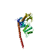
Mass: 18.015 Da / Num. of mol.: 36 / Source method: isolated from a natural source / Formula: H2O
Compound detailsPROBABLY INVOLVED IN ANCHORING OF MAJOR CYTOSKELETAL STRUCTURES TO THE PLASMA MEMBRANE.

-
Experimental details

-
Experiment

ExperimentMethod: X-RAY DIFFRACTION / Number of used crystals: 1

-
Sample preparation

CrystalDensity Matthews: 4.23 Å3/Da / Density % sol: 66 %
Crystal growpH: 7.5
Details: 25% ETHYLENE GLYCOL, 100MM HEPES PH7.5, 10-15 MG/ML PROTEIN, pH 7.50
Crystal grow
*PLUS
Temperature: 16 ℃ / Method: vapor diffusion, hanging drop
Components of the solutions
*PLUS
IDConc.Common nameCrystal-IDSol-ID
110-15 mg/mlprotein1drop
2100 mMHEPES1reservoir
325 %ethylene glycol1reservoir

-
Data collection

DiffractionMean temperature: 100 K
Diffraction sourceSource: SYNCHROTRON / Site: SRS / Beamline: PX9.6 / Wavelength: 0.87, 1.3
DetectorType: MARRESEARCH / Detector: CCD / Date: Oct 15, 1999
RadiationProtocol: MAD / Monochromatic (M) / Laue (L): M / Scattering type: x-ray
Radiation wavelength
IDWavelength (Å)Relative weight
10.871
21.31
ReflectionResolution: 2.7→37 Å / Num. obs: 19994 / % possible obs: 99.7 % / Observed criterion σ(I): 0 / Redundancy: 5.5 % / Biso Wilson estimate: 55.3 Å2 / Rmerge(I) obs: 0.046 / Rsym value: 0.046 / Net I/σ(I): 4.6
Reflection shellResolution: 2.7→2.85 Å / Redundancy: 5.5 % / Rmerge(I) obs: 0.246 / Mean I/σ(I) obs: 3 / Rsym value: 0.246 / % possible all: 99.9
Reflection
*PLUS
Lowest resolution: 37 Å / Num. measured all: 110406
Reflection shell
*PLUS
Highest resolution: 2.7 Å / % possible obs: 99.9 % / Num. unique obs: 2805 / Num. measured obs: 15646

-
Processing

Software
NameVersionClassification
MOSFLMdata reduction
SCALAdata scaling
SOLVEphasing
SHARPphasing
CNS1refinement
RefinementMethod to determine structure: MAD / Resolution: 2.7→37.12 Å / Rfactor Rfree error: 0.009 / Data cutoff high absF: 1200346.62 / Isotropic thermal model: RESTRAINED / Cross valid method: THROUGHOUT / σ(F): 0
RfactorNum. reflection% reflectionSelection details
Rfree0.297 1019 5.1 %RANDOM
Rwork0.254 ---
obs0.254 19994 99.7 %-
Solvent computationSolvent model: FLAT MODEL / Bsol: 38.5186 Å2 / ksol: 0.314713 e/Å3
Displacement parametersBiso mean: 73.5 Å2
Baniso -1Baniso -2Baniso -3
1--5.35 Å20 Å20 Å2
2---5.35 Å20 Å2
3---10.71 Å2
Refine analyze
FreeObs
Luzzati coordinate error0.55 Å0.45 Å
Luzzati d res low-5 Å
Luzzati sigma a0.63 Å0.48 Å
Refinement stepCycle: LAST / Resolution: 2.7→37.12 Å
ProteinNucleic acidLigandSolventTotal
Num. atoms2889 0 0 36 2925
Refine LS restraints
Refine-IDTypeDev idealDev ideal target
X-RAY DIFFRACTIONc_bond_d0.007
X-RAY DIFFRACTIONc_bond_d_na
X-RAY DIFFRACTIONc_bond_d_prot
X-RAY DIFFRACTIONc_angle_d
X-RAY DIFFRACTIONc_angle_d_na
X-RAY DIFFRACTIONc_angle_d_prot
X-RAY DIFFRACTIONc_angle_deg1.3
X-RAY DIFFRACTIONc_angle_deg_na
X-RAY DIFFRACTIONc_angle_deg_prot
X-RAY DIFFRACTIONc_dihedral_angle_d22.2
X-RAY DIFFRACTIONc_dihedral_angle_d_na
X-RAY DIFFRACTIONc_dihedral_angle_d_prot
X-RAY DIFFRACTIONc_improper_angle_d0.88
X-RAY DIFFRACTIONc_improper_angle_d_na
X-RAY DIFFRACTIONc_improper_angle_d_prot
X-RAY DIFFRACTIONc_mcbond_it1.5
X-RAY DIFFRACTIONc_mcangle_it2
X-RAY DIFFRACTIONc_scbond_it2
X-RAY DIFFRACTIONc_scangle_it2.5
LS refinement shellResolution: 2.7→2.87 Å / Rfactor Rfree error: 0.036 / Total num. of bins used: 6
RfactorNum. reflection% reflection
Rfree0.45 160 4.9 %
Rwork0.427 3077 -
obs--99.8 %
Xplor file
Refine-IDSerial noParam fileTopol file
X-RAY DIFFRACTION1PROTEIN_REP.PARAMPROTEIN.TOP
X-RAY DIFFRACTION2WATER_REP.PARAM
Software
*PLUS
Name: CNS / Version: 1 / Classification: refinement
Refine LS restraints
*PLUS
Refine-IDTypeDev ideal
X-RAY DIFFRACTIONc_dihedral_angle_d
X-RAY DIFFRACTIONc_dihedral_angle_deg22.2
X-RAY DIFFRACTIONc_improper_angle_d
X-RAY DIFFRACTIONc_improper_angle_deg0.88
LS refinement shell
*PLUS
Rfactor Rfree: 0.45 / Rfactor obs: 0.425

+
About Yorodumi

-
News

-
Feb 9, 2022. New format data for meta-information of EMDB entries

New format data for meta-information of EMDB entries

  • Version 3 of the EMDB header file is now the official format.
  • The previous official version 1.9 will be removed from the archive.

Related info.:EMDB header

External links:wwPDB to switch to version 3 of the EMDB data model

-
Aug 12, 2020. Covid-19 info

Covid-19 info

URL: https://pdbjlvh1.pdbj.org/emnavi/covid19.php

New page: Covid-19 featured information page in EM Navigator.

Related info.:Covid-19 info / Mar 5, 2020. Novel coronavirus structure data

+
Mar 5, 2020. Novel coronavirus structure data

Novel coronavirus structure data

Related info.:Yorodumi Speices / Aug 12, 2020. Covid-19 info

External links:COVID-19 featured content - PDBj / Molecule of the Month (242):Coronavirus Proteases

+
Jan 31, 2019. EMDB accession codes are about to change! (news from PDBe EMDB page)

EMDB accession codes are about to change! (news from PDBe EMDB page)

  • The allocation of 4 digits for EMDB accession codes will soon come to an end. Whilst these codes will remain in use, new EMDB accession codes will include an additional digit and will expand incrementally as the available range of codes is exhausted. The current 4-digit format prefixed with “EMD-” (i.e. EMD-XXXX) will advance to a 5-digit format (i.e. EMD-XXXXX), and so on. It is currently estimated that the 4-digit codes will be depleted around Spring 2019, at which point the 5-digit format will come into force.
  • The EM Navigator/Yorodumi systems omit the EMD- prefix.

Related info.:Q: What is EMD? / ID/Accession-code notation in Yorodumi/EM Navigator

External links:EMDB Accession Codes are Changing Soon! / Contact to PDBj

+
Jul 12, 2017. Major update of PDB

Major update of PDB

  • wwPDB released updated PDB data conforming to the new PDBx/mmCIF dictionary.
  • This is a major update changing the version number from 4 to 5, and with Remediation, in which all the entries are updated.
  • In this update, many items about electron microscopy experimental information are reorganized (e.g. em_software).
  • Now, EM Navigator and Yorodumi are based on the updated data.

External links:wwPDB Remediation / Enriched Model Files Conforming to OneDep Data Standards Now Available in the PDB FTP Archive

-
Yorodumi

Thousand views of thousand structures

  • Yorodumi is a browser for structure data from EMDB, PDB, SASBDB, etc.
  • This page is also the successor to EM Navigator detail page, and also detail information page/front-end page for Omokage search.
  • The word "yorodu" (or yorozu) is an old Japanese word meaning "ten thousand". "mi" (miru) is to see.

Related info.:EMDB / PDB / SASBDB / Comparison of 3 databanks / Yorodumi Search / Aug 31, 2016. New EM Navigator & Yorodumi / Yorodumi Papers / Jmol/JSmol / Function and homology information / Changes in new EM Navigator and Yorodumi

Read more