 Movie
Movie Controller

[English] 日本語
 Yorodumi
- PDB-2yvc: Crystal structure of the Radixin FERM domain complexed with the N... -
+ Open data

- Basic information
| Entry | Database: PDB / ID: 2yvc | ||||||
|---|---|---|---|---|---|---|---|
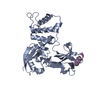
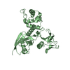
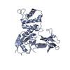
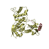
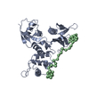
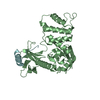
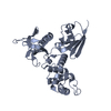
| Title | Crystal structure of the Radixin FERM domain complexed with the NEP cytoplasmic tail | ||||||
 Components Components |
| ||||||
 Keywords Keywords | CELL ADHESION / Protein-peptide complex | ||||||
| Function / homology |  Function and homology information Function and homology informationPhysiological factors / regulation of adherens junction organization / neuropeptide processing / neprilysin / creatinine metabolic process / stereocilium base / regulation of organelle assembly / microvillus assembly / establishment of protein localization to plasma membrane / amygdala development ...Physiological factors / regulation of adherens junction organization / neuropeptide processing / neprilysin / creatinine metabolic process / stereocilium base / regulation of organelle assembly / microvillus assembly / establishment of protein localization to plasma membrane / amygdala development / regulation of Rap protein signal transduction / Metabolism of Angiotensinogen to Angiotensins / positive regulation of early endosome to late endosome transport / oligopeptidase activity / Recycling pathway of L1 / exopeptidase activity / substance P catabolic process / cellular response to UV-A / positive regulation of protein localization to early endosome / amyloid-beta clearance by cellular catabolic process / regulation of postsynaptic neurotransmitter receptor diffusion trapping / peptide metabolic process / cell tip / hormone catabolic process / bradykinin catabolic process / barbed-end actin filament capping / stereocilium / cellular response to thyroid hormone stimulus / cardiolipin binding / apical protein localization / establishment of endothelial barrier / positive regulation of neurogenesis / cellular response to UV-B / cortical actin cytoskeleton / phosphatidylserine binding / cellular response to cytokine stimulus / neuron projection terminus / protein kinase A binding / microvillus / cleavage furrow / replicative senescence / amyloid-beta clearance / amyloid-beta metabolic process / brush border / lung development / positive regulation of G1/S transition of mitotic cell cycle / multicellular organism growth / angiotensin maturation / kidney development / metallocarboxypeptidase activity / ruffle / peptide binding / sensory perception of pain / Neutrophil degranulation / placenta development / protein catabolic process / cell adhesion molecule binding / T-tubule / hippocampus development / protein localization to plasma membrane / cell periphery / positive regulation of long-term synaptic potentiation / filopodium / adherens junction / establishment of protein localization / trans-Golgi network / protein processing / metalloendopeptidase activity / memory / response to estrogen / apical part of cell / metallopeptidase activity / positive regulation of protein catabolic process / peptidase activity / myelin sheath / regulation of cell shape / lamellipodium / synaptic vesicle / ATPase binding / presynapse / actin binding / cytoplasmic vesicle / midbody / endopeptidase activity / ciliary basal body / early endosome / learning or memory / apical plasma membrane / membrane raft / protein domain specific binding / axon / focal adhesion / neuronal cell body / centrosome / positive regulation of gene expression / synapse / dendrite / protein homodimerization activity / proteolysis / extracellular region Similarity search - Function | ||||||
| Biological species |  | ||||||
| Method |  X-RAY DIFFRACTION / SYNCHROTRON / MOLECULAR REPLACEMENT / Resolution: 3.2 Å X-RAY DIFFRACTION / SYNCHROTRON / MOLECULAR REPLACEMENT / Resolution: 3.2 Å | ||||||
 Authors Authors | Terawaki, S. / Kitano, K. / Hakoshima, T. | ||||||
 Citation Citation | Journal: J.Biol.Chem. / Year: 2007 Title: Structural basis for type II membrane protein binding by ERM proteins revealed by the radixin-neutral endopeptidase 24.11 (NEP) complex Authors: Terawaki, S. / Kitano, K. / Hakoshima, T. #1: Journal: Embo J. / Year: 2000Title: Structural basis of the membrane-targeting and unmasking mechanisms of the radixin FERM domain Authors: Hamada, K. / Shimizu, T. / Matsui, T. / Tsukita, S. / Hakoshima, T. #2: Journal: Embo J. / Year: 2003Title: Structural basis of adhesion-molecule recognition by ERM proteins revealed by the crystal structure of the radixin-ICAM-2 complex Authors: Hamada, K. / Shimizu, T. / Yonemura, S. / Tsukita, S. / Tsukita, S. / Hakoshima, T. #3: Journal: Structure / Year: 2006Title: Structural basis for NHERF recognition by ERM proteins Authors: Terawaki, S. / Maesaki, R. / Hakoshima, T. | ||||||
| History |
|
- Structure visualization
| Structure viewer | Molecule: MolmilJmol/JSmol |
|---|
- Downloads & links
-Download
| PDBx/mmCIF format | 2yvc.cif.gz | 357 KB | Display | PDBx/mmCIF format |
|---|---|---|---|---|
| PDB format | pdb2yvc.ent.gz | 294.4 KB | Display | PDB format |
| PDBx/mmJSON format | 2yvc.json.gz | Tree view | PDBx/mmJSON format | |
| Others |  Other downloads Other downloads |
-Validation report
| Arichive directory | https://data.pdbj.org/pub/pdb/validation_reports/yv/2yvcftp://data.pdbj.org/pub/pdb/validation_reports/yv/2yvc | HTTPS FTP |
|---|
-Related structure data
| Related structure data |  1gc7S S: Starting model for refinement |
|---|---|
| Similar structure data |


-Links
 PDBj
PDBj


- Assembly
| Deposited unit | 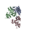
| ||||||||
|---|---|---|---|---|---|---|---|---|---|
| 1 | 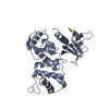
| ||||||||
| 2 | 
| ||||||||
| 3 | 
| ||||||||
| Unit cell |
|
-Components
| #1: Protein | Mass: 36924.559 Da / Num. of mol.: 3 / Fragment: FERM domain Source method: isolated from a genetically manipulated source Source: (gene. exp.)  #2: Protein/peptide | Mass: 2533.902 Da / Num. of mol.: 3 / Fragment: cytoplasmic tail / Source method: obtained synthetically / Details: This sequence occurs naturally in mouse. / References: UniProt: Q61391 |
|---|
-Experimental details
-Experiment
| Experiment | Method: X-RAY DIFFRACTION / Number of used crystals: 1 |
|---|
- Sample preparation
| Crystal | Density Matthews: 3.81 Å3/Da / Density % sol: 67.69 % |
|---|---|
| Crystal grow | Temperature: 277 K / Method: vapor diffusion, sitting drop / pH: 7 Details: 20% PEG3350, 0.2M DL-maleic acid, pH 7.0, VAPOR DIFFUSION, SITTING DROP, temperature 277K |
-Data collection
| Diffraction | Mean temperature: 100 K |
|---|---|
| Diffraction source | Source: SYNCHROTRON / Site: SPring-8  / Beamline: BL41XU / Wavelength: 1 Å / Beamline: BL41XU / Wavelength: 1 Å |
| Detector | Type: ADSC QUANTUM 315 / Detector: CCD / Date: Jun 28, 2005 |
| Radiation | Monochromator: rotated-inclined double-crystal / Protocol: SINGLE WAVELENGTH / Monochromatic (M) / Laue (L): M / Scattering type: x-ray |
| Radiation wavelength | Wavelength: 1 Å / Relative weight: 1 |
| Reflection | Resolution: 3.2→29.71 Å / Num. obs: 30656 / % possible obs: 99.7 % |
| Reflection shell | Resolution: 3.2→3.31 Å / % possible all: 97.2 |
- Processing
| Software |
| ||||||||||||||||||||||||||||||||||||||||||||||||||||||||||||||||||||||||||||||||||||||||||||||||||||||||||||||||||||||||||||||||||||||||||||||||||||||||||||||||||||||||||
|---|---|---|---|---|---|---|---|---|---|---|---|---|---|---|---|---|---|---|---|---|---|---|---|---|---|---|---|---|---|---|---|---|---|---|---|---|---|---|---|---|---|---|---|---|---|---|---|---|---|---|---|---|---|---|---|---|---|---|---|---|---|---|---|---|---|---|---|---|---|---|---|---|---|---|---|---|---|---|---|---|---|---|---|---|---|---|---|---|---|---|---|---|---|---|---|---|---|---|---|---|---|---|---|---|---|---|---|---|---|---|---|---|---|---|---|---|---|---|---|---|---|---|---|---|---|---|---|---|---|---|---|---|---|---|---|---|---|---|---|---|---|---|---|---|---|---|---|---|---|---|---|---|---|---|---|---|---|---|---|---|---|---|---|---|---|---|---|---|---|---|---|
| Refinement | Method to determine structure: MOLECULAR REPLACEMENT Starting model: PDB ENTRY 1GC7 Resolution: 3.2→29.71 Å / Cor.coef. Fo:Fc: 0.898 / Cor.coef. Fo:Fc free: 0.876 / SU B: 45.646 / SU ML: 0.346 / Cross valid method: THROUGHOUT / ESU R Free: 0.446 / Stereochemistry target values: MAXIMUM LIKELIHOOD / Details: HYDROGENS HAVE BEEN ADDED IN THE RIDING POSITIONS
| ||||||||||||||||||||||||||||||||||||||||||||||||||||||||||||||||||||||||||||||||||||||||||||||||||||||||||||||||||||||||||||||||||||||||||||||||||||||||||||||||||||||||||
| Solvent computation | Ion probe radii: 0.8 Å / Shrinkage radii: 0.8 Å / VDW probe radii: 1.4 Å / Solvent model: BABINET MODEL WITH MASK | ||||||||||||||||||||||||||||||||||||||||||||||||||||||||||||||||||||||||||||||||||||||||||||||||||||||||||||||||||||||||||||||||||||||||||||||||||||||||||||||||||||||||||
| Displacement parameters | Biso mean: 64.391 Å2
| ||||||||||||||||||||||||||||||||||||||||||||||||||||||||||||||||||||||||||||||||||||||||||||||||||||||||||||||||||||||||||||||||||||||||||||||||||||||||||||||||||||||||||
| Refinement step | Cycle: LAST / Resolution: 3.2→29.71 Å
| ||||||||||||||||||||||||||||||||||||||||||||||||||||||||||||||||||||||||||||||||||||||||||||||||||||||||||||||||||||||||||||||||||||||||||||||||||||||||||||||||||||||||||
| Refine LS restraints |
| ||||||||||||||||||||||||||||||||||||||||||||||||||||||||||||||||||||||||||||||||||||||||||||||||||||||||||||||||||||||||||||||||||||||||||||||||||||||||||||||||||||||||||
| LS refinement shell | Resolution: 3.195→3.277 Å / Total num. of bins used: 20
|
