- PDB-2d2q: Crystal structure of the dimerized radixin FERM domain -
+
Open data
ID or keywords:
Loading...
-
Basic information
Entry
Database: PDB / ID: 2d2q
Title
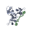
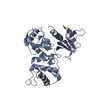
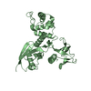
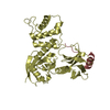
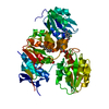
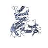
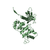
Crystal structure of the dimerized radixin FERM domain
Components
Radixin
Keywords
CELL ADHESION / Homo dimer / Masking
Function / homology
Function and homology information
regulation of adherens junction organization / stereocilium base / regulation of organelle assembly / microvillus assembly / establishment of protein localization to plasma membrane / positive regulation of early endosome to late endosome transport / regulation of Rap protein signal transduction / Recycling pathway of L1 / positive regulation of protein localization to early endosome / : ...regulation of adherens junction organization / stereocilium base / regulation of organelle assembly / microvillus assembly / establishment of protein localization to plasma membrane / positive regulation of early endosome to late endosome transport / regulation of Rap protein signal transduction / Recycling pathway of L1 / positive regulation of protein localization to early endosome / : / regulation of postsynaptic neurotransmitter receptor diffusion trapping / cell tip / barbed-end actin filament capping / stereocilium / cellular response to thyroid hormone stimulus / establishment of endothelial barrier / apical protein localization / cortical actin cytoskeleton / protein kinase A binding / microvillus / cleavage furrow / positive regulation of G1/S transition of mitotic cell cycle / cellular response to platelet-derived growth factor stimulus / ruffle / cell adhesion molecule binding / T-tubule / cell periphery / protein localization to plasma membrane / filopodium / adherens junction / establishment of protein localization / apical part of cell / positive regulation of protein catabolic process / regulation of cell shape / myelin sheath / lamellipodium / ATPase binding / actin binding / midbody / apical plasma membrane / protein domain specific binding / focal adhesion / positive regulation of gene expression / plasma membrane / cytosol Similarity search - Function
Moesin tail domain superfamily / Ezrin/radixin/moesin / Ezrin/radixin/moesin, C-terminal / ERM family, FERM domain C-lobe / Ezrin/radixin/moesin, alpha-helical domain / Ezrin/radixin/moesin family C terminal / Ezrin/radixin/moesin, alpha-helical domain / Acyl-CoA Binding Protein - #10 / Acyl-CoA Binding Protein / Ezrin/radixin/moesin-like ...Moesin tail domain superfamily / Ezrin/radixin/moesin / Ezrin/radixin/moesin, C-terminal / ERM family, FERM domain C-lobe / Ezrin/radixin/moesin, alpha-helical domain / Ezrin/radixin/moesin family C terminal / Ezrin/radixin/moesin, alpha-helical domain / Acyl-CoA Binding Protein - #10 / Acyl-CoA Binding Protein / Ezrin/radixin/moesin-like / FERM, C-terminal PH-like domain / FERM C-terminal PH-like domain / FERM C-terminal PH-like domain / FERM, N-terminal / FERM N-terminal domain / FERM domain signature 1. / FERM conserved site / FERM domain signature 2. / FERM central domain / FERM/acyl-CoA-binding protein superfamily / Pleckstrin-homology domain (PH domain)/Phosphotyrosine-binding domain (PTB) / PH-domain like / FERM central domain / FERM superfamily, second domain / FERM domain / FERM domain profile. / Band 4.1 domain / Band 4.1 homologues / Phosphatidylinositol 3-kinase Catalytic Subunit; Chain A, domain 1 / Ubiquitin-like (UB roll) / PH-like domain superfamily / Roll / Ubiquitin-like domain superfamily / Roll / Up-down Bundle / Mainly Beta / Mainly Alpha / Alpha Beta Similarity search - Domain/homology
In the structure databanks used in Yorodumi, some data are registered as the other names, "COVID-19 virus" and "2019-nCoV". Here are the details of the virus and the list of structure data.
Jan 31, 2019. EMDB accession codes are about to change! (news from PDBe EMDB page)
EMDB accession codes are about to change! (news from PDBe EMDB page)
The allocation of 4 digits for EMDB accession codes will soon come to an end. Whilst these codes will remain in use, new EMDB accession codes will include an additional digit and will expand incrementally as the available range of codes is exhausted. The current 4-digit format prefixed with “EMD-” (i.e. EMD-XXXX) will advance to a 5-digit format (i.e. EMD-XXXXX), and so on. It is currently estimated that the 4-digit codes will be depleted around Spring 2019, at which point the 5-digit format will come into force.
The EM Navigator/Yorodumi systems omit the EMD- prefix.
Related info.:Q: What is EMD? / ID/Accession-code notation in Yorodumi/EM Navigator
Yorodumi is a browser for structure data from EMDB, PDB, SASBDB, etc.
This page is also the successor to EM Navigator detail page, and also detail information page/front-end page for Omokage search.
The word "yorodu" (or yorozu) is an old Japanese word meaning "ten thousand". "mi" (miru) is to see.
Related info.:EMDB / PDB / SASBDB / Comparison of 3 databanks / Yorodumi Search / Aug 31, 2016. New EM Navigator & Yorodumi / Yorodumi Papers / Jmol/JSmol / Function and homology information / Changes in new EM Navigator and Yorodumi
 Movie
Movie Controller


 Open data
 Basic information
 Components
 Keywords
 Function and homology information
 X-RAY DIFFRACTION /
  Authors
 Citation
 Structure visualization
 Downloads & links
 Other downloads
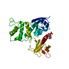


 PDBj
PDBj

 Assembly
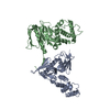

 Sample preparation
 / Beamline: BL44XU / Wavelength: 0.9 Å
 Processing