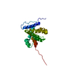
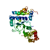
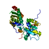
[English] 日本語
Yorodumi
- PDB-2y6w: Structure of a Bcl-w dimer -

+
Open data


ID or keywords:

Loading...

-
Basic information

Entry
Database: PDB / ID: 2y6w
TitleStructure of a Bcl-w dimer
ComponentsBCL-2-LIKE PROTEIN 2
KeywordsAPOPTOSIS
Function / homology
Function and homology information


negative regulation of mitochondrial membrane permeability / Sertoli cell proliferation / BH domain binding / Bcl-2 family protein complex / negative regulation of release of cytochrome c from mitochondria / cellular response to glycine / negative regulation of intrinsic apoptotic signaling pathway / extrinsic apoptotic signaling pathway in absence of ligand / cellular response to estradiol stimulus / response to ischemia ...negative regulation of mitochondrial membrane permeability / Sertoli cell proliferation / BH domain binding / Bcl-2 family protein complex / negative regulation of release of cytochrome c from mitochondria / cellular response to glycine / negative regulation of intrinsic apoptotic signaling pathway / extrinsic apoptotic signaling pathway in absence of ligand / cellular response to estradiol stimulus / response to ischemia / cellular response to amyloid-beta / intrinsic apoptotic signaling pathway in response to DNA damage / disordered domain specific binding / spermatogenesis / regulation of apoptotic process / mitochondrial outer membrane / protein heterodimerization activity / protein-containing complex binding / negative regulation of apoptotic process / protein homodimerization activity / identical protein binding / cytosol
Similarity search - Function
Apoptosis regulator, Bcl-W / Apoptosis regulator, Bcl-2, BH4 motif, conserved site / Apoptosis regulator, Bcl-2 family BH4 motif signature. / Apoptosis regulator, Bcl-2 protein, BH4 / Bcl-2 homology region 4 / Apoptosis regulator, Bcl-2 family BH4 motif profile. / BH4 Bcl-2 homology region 4 / Apoptosis regulator, Bcl-2, BH1 motif, conserved site / Apoptosis regulator, Bcl-2 family BH1 motif signature. / Apoptosis regulator, Bcl-2, BH2 motif, conserved site ...Apoptosis regulator, Bcl-W / Apoptosis regulator, Bcl-2, BH4 motif, conserved site / Apoptosis regulator, Bcl-2 family BH4 motif signature. / Apoptosis regulator, Bcl-2 protein, BH4 / Bcl-2 homology region 4 / Apoptosis regulator, Bcl-2 family BH4 motif profile. / BH4 Bcl-2 homology region 4 / Apoptosis regulator, Bcl-2, BH1 motif, conserved site / Apoptosis regulator, Bcl-2 family BH1 motif signature. / Apoptosis regulator, Bcl-2, BH2 motif, conserved site / Apoptosis regulator, Bcl-2 family BH2 motif signature. / BCL (B-Cell lymphoma); contains BH1, BH2 regions / Bcl-2 family / Bcl-2, Bcl-2 homology region 1-3 / Bcl2-like / Apoptosis regulator proteins, Bcl-2 family / BCL2-like apoptosis inhibitors family profile. / Bcl-2-like superfamily
Similarity search - Domain/homology
DI(HYDROXYETHYL)ETHER / TRIETHYLENE GLYCOL / Bcl-2-like protein 2
Similarity search - Component
Biological speciesHOMO SAPIENS (human)
MethodX-RAY DIFFRACTION / SYNCHROTRON / MOLECULAR REPLACEMENT / Resolution: 2 Å
AuthorsLee, E.F. / Evangelista, M. / Pettikiriarachchi, A. / Dogovski, C. / Perugini, M.A. / Colman, P.M. / Fairlie, W.D.
CitationJournal: Structure / Year: 2011
Title: Crystal Structure of a Bcl-W Domain-Swapped Dimer: Implications for the Function of Bcl-2 Family Proteins.
Authors: Lee, E.F. / Dewson, G. / Smith, B.J. / Evangelista, M. / Pettikiriarachchi, A. / Dogovski, C. / Perugini, M.A. / Colman, P.M. / Fairlie, W.D.
History
DepositionJan 27, 2011Deposition site: PDBE / Processing site: PDBE
Revision 1.0Oct 26, 2011Provider: repository / Type: Initial release
Revision 1.1Dec 20, 2023Group: Data collection / Database references ...Data collection / Database references / Derived calculations / Other / Refinement description
Category: chem_comp_atom / chem_comp_bond ...chem_comp_atom / chem_comp_bond / database_2 / pdbx_database_status / pdbx_initial_refinement_model / struct_site
Item: _database_2.pdbx_DOI / _database_2.pdbx_database_accession ..._database_2.pdbx_DOI / _database_2.pdbx_database_accession / _pdbx_database_status.status_code_sf / _struct_site.pdbx_auth_asym_id / _struct_site.pdbx_auth_comp_id / _struct_site.pdbx_auth_seq_id

-
Structure visualization

Structure viewerMolecule:
MolmilJmol/JSmol

Downloads & links

-
Assembly

Deposited unit
A: BCL-2-LIKE PROTEIN 2
B: BCL-2-LIKE PROTEIN 2
hetero molecules


Theoretical massNumber of molelcules
Total (without water)39,6928
Polymers39,0112
Non-polymers6816
Water2,774154
1


  • Idetical with deposited unit
  • defined by author&software
TypeNameSymmetry operationNumber
identity operation1_555x,y,z1
Buried area5490 Å2
ΔGint-12.8 kcal/mol
Surface area14310 Å2
MethodPISA
Unit cell
Length a, b, c (Å)76.227, 76.227, 112.847
Angle α, β, γ (deg.)90.00, 90.00, 90.00
Int Tables number96
Space group name H-MP43212
Components on special symmetry positions
IDModelComponents
11B-2043-

HOH

-
Components

#1: Protein BCL-2-LIKE PROTEIN 2 / BCL-W / BCL2-L-2 / APOPTOSIS REGULATOR BCL-W


Mass: 19505.693 Da / Num. of mol.: 2 / Fragment: C-TERMINAL TRUNCATION, RESIDUES 1-164 / Mutation: YES
Source method: isolated from a genetically manipulated source
Source: (gene. exp.) HOMO SAPIENS (human) / Production host: ESCHERICHIA COLI (E. coli) / Strain (production host): BL21 / References: UniProt: Q92843
#2: Chemical
ChemComp-PEG / DI(HYDROXYETHYL)ETHER


Mass: 106.120 Da / Num. of mol.: 5 / Source method: obtained synthetically / Formula: C4H10O3
#3: Chemical ChemComp-PGE / TRIETHYLENE GLYCOL


Mass: 150.173 Da / Num. of mol.: 1 / Source method: obtained synthetically / Formula: C6H14O4
#4: Water ChemComp-HOH / water


Mass: 18.015 Da / Num. of mol.: 154 / Source method: isolated from a natural source / Formula: H2O
Compound details
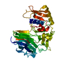
ENGINEERED RESIDUE IN CHAIN A, CYS 29 TO SER ENGINEERED RESIDUE IN CHAIN A, ALA 128 TO GLU ...ENGINEERED RESIDUE IN CHAIN A, CYS 29 TO SER ENGINEERED RESIDUE IN CHAIN A, ALA 128 TO GLU ENGINEERED RESIDUE IN CHAIN B, CYS 29 TO SER ENGINEERED RESIDUE IN CHAIN B, ALA 128 TO GLU
Sequence detailsC29S AND A128E MUTATIONS, TRUNCATION OF LAST 29 AMINO ACID RESIDUES.

-
Experimental details

-
Experiment

ExperimentMethod: X-RAY DIFFRACTION / Number of used crystals: 1

-
Sample preparation

CrystalDensity Matthews: 2.1 Å3/Da / Density % sol: 41.46 % / Description: NONE
Crystal growpH: 6.5
Details: 5% (W/V) PEG 3000, 40% (W/V) PEG 400, 0.1M MES PH 6.5.

-
Data collection

DiffractionMean temperature: 100 K
Diffraction sourceSource: SYNCHROTRON / Site: SLS / Beamline: X06DA / Wavelength: 1
DetectorType: MARMOSAIC 225 mm CCD / Detector: CCD / Date: Aug 4, 2008
RadiationProtocol: SINGLE WAVELENGTH / Monochromatic (M) / Laue (L): M / Scattering type: x-ray
Radiation wavelengthWavelength: 1 Å / Relative weight: 1
ReflectionResolution: 2→49 Å / Num. obs: 23159 / % possible obs: 99.9 % / Observed criterion σ(I): -3 / Redundancy: 11.8 % / Biso Wilson estimate: 36.92 Å2 / Rmerge(I) obs: 0.08 / Net I/σ(I): 20.5
Reflection shellResolution: 2→2.09 Å / Redundancy: 11.8 % / Rmerge(I) obs: 1.31 / Mean I/σ(I) obs: 2.2 / % possible all: 99.5

-
Processing

Software
NameVersionClassification
PHENIX(PHENIX.REFINE)refinement
HKL-2000data reduction
HKL-2000data scaling
PHASERphasing
RefinementMethod to determine structure: MOLECULAR REPLACEMENT
Starting model: PDB ENTRY 1PQ0
Resolution: 2→48.637 Å / SU ML: 1.36 / σ(F): 2 / Phase error: 22.13 / Stereochemistry target values: ML
RfactorNum. reflection% reflection
Rfree0.2442 1157 5 %
Rwork0.1989 --
obs0.2011 23135 99.93 %
Solvent computationShrinkage radii: 0.9 Å / VDW probe radii: 1.11 Å / Solvent model: FLAT BULK SOLVENT MODEL / Bsol: 64.004 Å2 / ksol: 0.391 e/Å3
Displacement parameters
Baniso -1Baniso -2Baniso -3
1-1.8368 Å20 Å20 Å2
2--1.8368 Å20 Å2
3----3.6736 Å2
Refinement stepCycle: LAST / Resolution: 2→48.637 Å
ProteinNucleic acidLigandSolventTotal
Num. atoms2270 0 45 154 2469
Refine LS restraints
Refine-IDTypeDev idealNumber
X-RAY DIFFRACTIONf_bond_d0.0082371
X-RAY DIFFRACTIONf_angle_d1.0483198
X-RAY DIFFRACTIONf_dihedral_angle_d17.309837
X-RAY DIFFRACTIONf_chiral_restr0.065323
X-RAY DIFFRACTIONf_plane_restr0.005424
LS refinement shell
Resolution (Å)Rfactor RfreeNum. reflection RfreeRfactor RworkNum. reflection RworkRefine-ID% reflection obs (%)
2.0001-2.09110.27691410.24152678X-RAY DIFFRACTION100
2.0911-2.20130.2611420.21712701X-RAY DIFFRACTION100
2.2013-2.33930.26631420.21132704X-RAY DIFFRACTION100
2.3393-2.51990.26291420.20442707X-RAY DIFFRACTION100
2.5199-2.77340.29151440.20442723X-RAY DIFFRACTION100
2.7734-3.17470.26531450.21052758X-RAY DIFFRACTION100
3.1747-3.99950.23311460.18672778X-RAY DIFFRACTION100
3.9995-48.65160.21241550.18542929X-RAY DIFFRACTION100

+
About Yorodumi

-
News

-
Feb 9, 2022. New format data for meta-information of EMDB entries

New format data for meta-information of EMDB entries

  • Version 3 of the EMDB header file is now the official format.
  • The previous official version 1.9 will be removed from the archive.

Related info.:EMDB header

External links:wwPDB to switch to version 3 of the EMDB data model

-
Aug 12, 2020. Covid-19 info

Covid-19 info

URL: https://pdbjlvh1.pdbj.org/emnavi/covid19.php

New page: Covid-19 featured information page in EM Navigator.

Related info.:Covid-19 info / Mar 5, 2020. Novel coronavirus structure data

+
Mar 5, 2020. Novel coronavirus structure data

Novel coronavirus structure data

Related info.:Yorodumi Speices / Aug 12, 2020. Covid-19 info

External links:COVID-19 featured content - PDBj / Molecule of the Month (242):Coronavirus Proteases

+
Jan 31, 2019. EMDB accession codes are about to change! (news from PDBe EMDB page)

EMDB accession codes are about to change! (news from PDBe EMDB page)

  • The allocation of 4 digits for EMDB accession codes will soon come to an end. Whilst these codes will remain in use, new EMDB accession codes will include an additional digit and will expand incrementally as the available range of codes is exhausted. The current 4-digit format prefixed with “EMD-” (i.e. EMD-XXXX) will advance to a 5-digit format (i.e. EMD-XXXXX), and so on. It is currently estimated that the 4-digit codes will be depleted around Spring 2019, at which point the 5-digit format will come into force.
  • The EM Navigator/Yorodumi systems omit the EMD- prefix.

Related info.:Q: What is EMD? / ID/Accession-code notation in Yorodumi/EM Navigator

External links:EMDB Accession Codes are Changing Soon! / Contact to PDBj

+
Jul 12, 2017. Major update of PDB

Major update of PDB

  • wwPDB released updated PDB data conforming to the new PDBx/mmCIF dictionary.
  • This is a major update changing the version number from 4 to 5, and with Remediation, in which all the entries are updated.
  • In this update, many items about electron microscopy experimental information are reorganized (e.g. em_software).
  • Now, EM Navigator and Yorodumi are based on the updated data.

External links:wwPDB Remediation / Enriched Model Files Conforming to OneDep Data Standards Now Available in the PDB FTP Archive

-
Yorodumi

Thousand views of thousand structures

  • Yorodumi is a browser for structure data from EMDB, PDB, SASBDB, etc.
  • This page is also the successor to EM Navigator detail page, and also detail information page/front-end page for Omokage search.
  • The word "yorodu" (or yorozu) is an old Japanese word meaning "ten thousand". "mi" (miru) is to see.

Related info.:EMDB / PDB / SASBDB / Comparison of 3 databanks / Yorodumi Search / Aug 31, 2016. New EM Navigator & Yorodumi / Yorodumi Papers / Jmol/JSmol / Function and homology information / Changes in new EM Navigator and Yorodumi

Read more