 Movie
Movie Controller

[English] 日本語
 Yorodumi
- PDB-2egu: Crystal structure of O-acetylserine sulfhydrase from Geobacillus ... -
+ Open data

- Basic information
| Entry | Database: PDB / ID: 2egu | ||||||
|---|---|---|---|---|---|---|---|
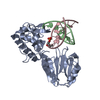
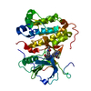
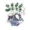
| Title | Crystal structure of O-acetylserine sulfhydrase from Geobacillus kaustophilus HTA426 | ||||||
 Components Components | Cysteine synthase | ||||||
 Keywords Keywords | TRANSFERASE / O-acetylserine sulfhydrase / Structural Genomics / NPPSFA / National Project on Protein Structural and Functional Analyses / RIKEN Structural Genomics/Proteomics Initiative / RSGI | ||||||
| Function / homology |  Function and homology information Function and homology information | ||||||
| Biological species |  Geobacillus kaustophilus (bacteria) Geobacillus kaustophilus (bacteria) | ||||||
| Method |  X-RAY DIFFRACTION / SYNCHROTRON / MOLECULAR REPLACEMENT / Resolution: 1.9 Å X-RAY DIFFRACTION / SYNCHROTRON / MOLECULAR REPLACEMENT / Resolution: 1.9 Å | ||||||
 Authors Authors | Rehse, P.H. / Yokoyama, S. / RIKEN Structural Genomics/Proteomics Initiative (RSGI) | ||||||
 Citation Citation | Journal: To be Published Title: Crystal structure of O-acetylserine sulfhydrase from Geobacillus kaustophilus HTA426 Authors: Rehse, P.H. / Yokoyama, S. | ||||||
| History |
|
- Structure visualization
| Structure viewer | Molecule: MolmilJmol/JSmol |
|---|
- Downloads & links
-Download
| PDBx/mmCIF format | 2egu.cif.gz | 68.9 KB | Display | PDBx/mmCIF format |
|---|---|---|---|---|
| PDB format | pdb2egu.ent.gz | 49.8 KB | Display | PDB format |
| PDBx/mmJSON format | 2egu.json.gz | Tree view | PDBx/mmJSON format | |
| Others |  Other downloads Other downloads |
-Validation report
| Arichive directory | https://data.pdbj.org/pub/pdb/validation_reports/eg/2eguftp://data.pdbj.org/pub/pdb/validation_reports/eg/2egu | HTTPS FTP |
|---|
-Related structure data
| Related structure data | 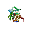 1z7wS S: Starting model for refinement |
|---|---|
| Similar structure data | |
| Other databases |


-Links
 PDBj
PDBj
- Assembly
| Deposited unit | 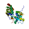
| |||||||||
|---|---|---|---|---|---|---|---|---|---|---|
| 1 |
| |||||||||
| 2 | 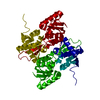
| |||||||||
| 3 | 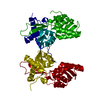
| |||||||||
| Unit cell |
| |||||||||
| Components on special symmetry positions |
|
-Components
| #1: Protein | Mass: 32555.275 Da / Num. of mol.: 1 Source method: isolated from a genetically manipulated source Source: (gene. exp.) Geobacillus kaustophilus (bacteria) / Strain: HTA426 / Plasmid: pET-15B / Production host: |
|---|---|
| #2: Water | ChemComp-HOH /  Mass: 18.015 Da / Num. of mol.: 246 / Source method: isolated from a natural source / Formula: H2O Mass: 18.015 Da / Num. of mol.: 246 / Source method: isolated from a natural source / Formula: H2O |
-Experimental details
-Experiment
| Experiment | Method: X-RAY DIFFRACTION / Number of used crystals: 1 |
|---|
- Sample preparation
| Crystal | Density Matthews: 2.55 Å3/Da / Density % sol: 51.68 % |
|---|---|
| Crystal grow | Temperature: 293 K / Method: vapor diffusion, sitting drop / pH: 6.3 Details: 100mM MES-NaOH, 50mM MgCl2, 27.5% PEG 4000, pH 6.3, VAPOR DIFFUSION, SITTING DROP, temperature 293K |
-Data collection
| Diffraction | Mean temperature: 100 K |
|---|---|
| Diffraction source | Source: SYNCHROTRON / Site: SPring-8  / Beamline: BL45XU / Wavelength: 0.9794 Å / Beamline: BL45XU / Wavelength: 0.9794 Å |
| Detector | Type: RIGAKU RAXIS V / Detector: IMAGE PLATE / Date: Dec 16, 2006 / Details: mirrors |
| Radiation | Monochromator: Si (111) / Protocol: SINGLE WAVELENGTH / Monochromatic (M) / Laue (L): M / Scattering type: x-ray |
| Radiation wavelength | Wavelength: 0.9794 Å / Relative weight: 1 |
| Reflection | Resolution: 1.9→40 Å / Num. all: 27260 / Num. obs: 27236 / % possible obs: 99.9 % / Observed criterion σ(F): 0 / Observed criterion σ(I): 0 / Redundancy: 14.3 % / Rmerge(I) obs: 0.107 / Net I/σ(I): 36.86 |
| Reflection shell | Resolution: 1.9→1.97 Å / Redundancy: 14.6 % / Rmerge(I) obs: 0.529 / Mean I/σ(I) obs: 5.22 / Num. unique all: 2654 / % possible all: 100 |
- Processing
| Software |
| ||||||||||||||||||||||||||||
|---|---|---|---|---|---|---|---|---|---|---|---|---|---|---|---|---|---|---|---|---|---|---|---|---|---|---|---|---|---|
| Refinement | Method to determine structure: MOLECULAR REPLACEMENT Starting model: 1Z7W.pdb Resolution: 1.9→39.81 Å / Isotropic thermal model: anisotropic / Cross valid method: THROUGHOUT / σ(F): 0 / σ(I): 0 / Stereochemistry target values: Engh & Huber
| ||||||||||||||||||||||||||||
| Solvent computation | Bsol: 35.998 Å2 | ||||||||||||||||||||||||||||
| Displacement parameters | Biso mean: 28.574 Å2
| ||||||||||||||||||||||||||||
| Refine analyze |
| ||||||||||||||||||||||||||||
| Refinement step | Cycle: LAST / Resolution: 1.9→39.81 Å
| ||||||||||||||||||||||||||||
| Refine LS restraints |
| ||||||||||||||||||||||||||||
| LS refinement shell | Resolution: 1.9→1.97 Å
| ||||||||||||||||||||||||||||
| Xplor file |
|
