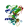
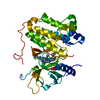
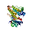
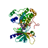
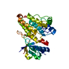
Entry Database : PDB / ID : 5gmpTitle Crystal structure of EGFR 696-1022 T790M in complex with XTF-262 Epidermal growth factor receptor Keywords / / / / Function / homology Function Domain/homology Component
/ / / / / / / / / / / / / / / / / / / / / / / / / / / / / / / / / / / / / / / / / / / / / / / / / / / / / / / / / / / / / / / / / / / / / / / / / / / / / / / / / / / / / / / / / / / / / / / / / / / / / / / / / / / / / / / / / / / / / / / / / / / / / / / / / / / / / / / / / / / / / / / / / / / / / Biological species Homo sapiens (human)Method / / / Resolution : 2.797 Å Authors Yan, X.E. / Yun, C.H. Journal : Eur J Med Chem / Year : 2017Title : A structure-guided optimization of pyrido[2,3-d]pyrimidin-7-ones as selective inhibitors of EGFR(L858R/T790M) mutant with improved pharmacokinetic properties.Authors : Yu, L. / Huang, M. / Xu, T. / Tong, L. / Yan, X.E. / Zhang, Z. / Xu, Y. / Yun, C. / Xie, H. / Ding, K. / Lu, X. History Deposition Jul 14, 2016 Deposition site / Processing site Revision 1.0 Jun 28, 2017 Provider / Type Revision 1.1 Oct 4, 2017 Group / Category / Item Revision 1.2 Nov 8, 2023 Group / Database references / Refinement descriptionCategory chem_comp_atom / chem_comp_bond ... chem_comp_atom / chem_comp_bond / database_2 / pdbx_initial_refinement_model Item / _database_2.pdbx_database_accessionRevision 1.3 Oct 16, 2024 Group / Category / pdbx_modification_feature
Show all Show less
 Movie
Movie Controller


 Open data
 Basic information
 Components
 Keywords
 Function and homology information
 Homo sapiens (human)
 X-RAY DIFFRACTION /
  Authors
 Citation
 Structure visualization
 Downloads & links
 Other downloads


 PDBj
PDBj


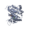
 Assembly
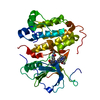

 Spodoptera frugiperda (fall armyworm)
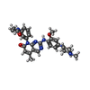
 Mass: 525.602 Da / Num. of mol.: 1 / Source method: obtained synthetically / Formula: C29H31N7O3
 Mass: 18.015 Da / Num. of mol.: 31 / Source method: isolated from a natural source / Formula: H2O
 Sample preparation
 / Beamline: BL18U1 / Wavelength: 0.97776 Å
 Processing