- PDB-3pvp: Structure of Mycobacterium tuberculosis DnaA-DBD in complex with ... -
+
Open data
ID or keywords:
Loading...
-
Basic information
Entry
Database: PDB / ID: 3pvp
Title
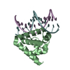
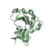
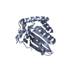
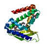
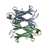
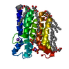
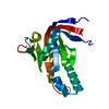
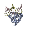
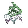
Structure of Mycobacterium tuberculosis DnaA-DBD in complex with box2 DNA
Components
Chromosomal replication initiator protein dnaA
DNA (5'-D(*CP*GP*TP*TP*AP*TP*CP*CP*AP*CP*AP*AP*C)-3')
DNA (5'-D(*GP*TP*TP*GP*TP*GP*GP*AP*TP*AP*AP*CP*G)-3')
Keywords
DNA BINDING PROTEIN/DNA / Helical / DNA replication / DNA binding / DnaA-box / DNA BINDING PROTEIN-DNA complex
Function / homology
Function and homology information
regulation of DNA replication / DNA replication origin binding / DNA replication initiation / lipid binding / ATP binding / plasma membrane / cytoplasm Similarity search - Function
A: Chromosomal replication initiator protein dnaA B: Chromosomal replication initiator protein dnaA C: DNA (5'-D(*CP*GP*TP*TP*AP*TP*CP*CP*AP*CP*AP*AP*C)-3') D: DNA (5'-D(*GP*TP*TP*GP*TP*GP*GP*AP*TP*AP*AP*CP*G)-3') E: DNA (5'-D(*CP*GP*TP*TP*AP*TP*CP*CP*AP*CP*AP*AP*C)-3') F: DNA (5'-D(*GP*TP*TP*GP*TP*GP*GP*AP*TP*AP*AP*CP*G)-3')
A: Chromosomal replication initiator protein dnaA C: DNA (5'-D(*CP*GP*TP*TP*AP*TP*CP*CP*AP*CP*AP*AP*C)-3') D: DNA (5'-D(*GP*TP*TP*GP*TP*GP*GP*AP*TP*AP*AP*CP*G)-3')
B: Chromosomal replication initiator protein dnaA E: DNA (5'-D(*CP*GP*TP*TP*AP*TP*CP*CP*AP*CP*AP*AP*C)-3') F: DNA (5'-D(*GP*TP*TP*GP*TP*GP*GP*AP*TP*AP*AP*CP*G)-3')
Mass: 18.015 Da / Num. of mol.: 84 / Source method: isolated from a natural source / Formula: H2O
-
Experimental details
-
Experiment
Experiment
Method: X-RAY DIFFRACTION / Number of used crystals: 1
-
Sample preparation
Crystal
Density Matthews: 3.06 Å3/Da / Density % sol: 59.78 %
Crystal grow
Temperature: 295 K / Method: vapor diffusion, hanging drop / pH: 8 Details: 20 mM Tris, 2 mM MgCl2, 50 mM NaCl, 1200 mM NH4SO4, 2-5% glycerol, pH 8.0, VAPOR DIFFUSION, HANGING DROP, temperature 295K
Type: MARMOSAIC 300 mm CCD / Detector: CCD / Date: Dec 6, 2007
Radiation
Protocol: SINGLE WAVELENGTH / Monochromatic (M) / Laue (L): M / Scattering type: x-ray
Radiation wavelength
Wavelength: 1 Å / Relative weight: 1
Reflection
Resolution: 2.2→50 Å / Num. obs: 21973 / % possible obs: 87.1 % / Observed criterion σ(I): 2
-
Processing
Software
Name
Version
Classification
HKL-2000
datacollection
PHASER
phasing
REFMAC
5.5.0088
refinement
HKL-2000
datareduction
HKL-2000
datascaling
Refinement
Method to determine structure: MOLECULAR REPLACEMENT / Resolution: 2.3→35 Å / Cor.coef. Fo:Fc: 0.948 / Cor.coef. Fo:Fc free: 0.945 / SU ML: 0.181 / Cross valid method: THROUGHOUT / ESU R Free: 0.215 / Stereochemistry target values: MAXIMUM LIKELIHOOD / Details: HYDROGENS HAVE BEEN ADDED IN THE RIDING POSITIONS
Rfactor
Num. reflection
% reflection
Selection details
Rfree
0.23821
1034
5.2 %
RANDOM
Rwork
0.22094
-
-
-
all
0.222
20594
-
-
obs
0.22184
18916
91.85 %
-
Solvent computation
Ion probe radii: 0.8 Å / Shrinkage radii: 0.8 Å / VDW probe radii: 1.4 Å / Solvent model: MASK
Refinement step
Cycle: LAST / Resolution: 2.3→35 Å
Protein
Nucleic acid
Ligand
Solvent
Total
Num. atoms
1516
1054
0
84
2654
Refine LS restraints
Refine-ID
Type
Dev ideal
Dev ideal target
Number
X-RAY DIFFRACTION
r_bond_refined_d
0.005
0.021
2714
X-RAY DIFFRACTION
r_bond_other_d
X-RAY DIFFRACTION
r_angle_refined_deg
0.924
2.449
3874
X-RAY DIFFRACTION
r_angle_other_deg
X-RAY DIFFRACTION
r_dihedral_angle_1_deg
4.03
5
189
X-RAY DIFFRACTION
r_dihedral_angle_2_deg
31.446
21.429
70
X-RAY DIFFRACTION
r_dihedral_angle_3_deg
16.142
15
293
X-RAY DIFFRACTION
r_dihedral_angle_4_deg
16.294
15
22
X-RAY DIFFRACTION
r_chiral_restr
X-RAY DIFFRACTION
r_gen_planes_refined
0.003
0.02
1670
X-RAY DIFFRACTION
r_gen_planes_other
X-RAY DIFFRACTION
r_nbd_refined
X-RAY DIFFRACTION
r_nbd_other
X-RAY DIFFRACTION
r_nbtor_refined
X-RAY DIFFRACTION
r_nbtor_other
X-RAY DIFFRACTION
r_xyhbond_nbd_refined
X-RAY DIFFRACTION
r_xyhbond_nbd_other
X-RAY DIFFRACTION
r_metal_ion_refined
X-RAY DIFFRACTION
r_metal_ion_other
X-RAY DIFFRACTION
r_symmetry_vdw_refined
X-RAY DIFFRACTION
r_symmetry_vdw_other
X-RAY DIFFRACTION
r_symmetry_hbond_refined
X-RAY DIFFRACTION
r_symmetry_hbond_other
X-RAY DIFFRACTION
r_symmetry_metal_ion_refined
X-RAY DIFFRACTION
r_symmetry_metal_ion_other
X-RAY DIFFRACTION
r_mcbond_it
X-RAY DIFFRACTION
r_mcbond_other
X-RAY DIFFRACTION
r_mcangle_it
X-RAY DIFFRACTION
r_scbond_it
X-RAY DIFFRACTION
r_scangle_it
X-RAY DIFFRACTION
r_rigid_bond_restr
X-RAY DIFFRACTION
r_sphericity_free
X-RAY DIFFRACTION
r_sphericity_bonded
LS refinement shell
Resolution: 2.3→2.36 Å / Total num. of bins used: 20
Rfactor
Num. reflection
% reflection
Rfree
0.346
44
-
Rwork
0.313
919
-
obs
-
-
61.07 %
Refinement TLS params.
Method: refined / Refine-ID: X-RAY DIFFRACTION
ID
L11 (°2)
L12 (°2)
L13 (°2)
L22 (°2)
L23 (°2)
L33 (°2)
S11 (Å °)
S12 (Å °)
S13 (Å °)
S21 (Å °)
S22 (Å °)
S23 (Å °)
S31 (Å °)
S32 (Å °)
S33 (Å °)
T11 (Å2)
T12 (Å2)
T13 (Å2)
T22 (Å2)
T23 (Å2)
T33 (Å2)
Origin x (Å)
Origin y (Å)
Origin z (Å)
1
1.4641
0.9342
0.4871
1.3791
-0.4783
1.258
-0.077
0.0664
0.0307
-0.0676
-0.005
0.0303
0.0351
0.0791
0.082
0.414
0.0019
0.0148
0.4332
0.0184
0.1706
-34.879
24.744
22.425
2
1.9
0.0024
0.3775
1.4314
0.567
1.5124
-0.0903
0.2073
0.1571
-0.157
0.0255
0.0068
-0.1555
0.0983
0.0649
0.434
-0.0063
-0.0269
0.438
0.0301
0.1753
-33.586
44.34
10.636
3
0.6094
0.8831
-1.3373
1.5074
-2.3628
3.7603
-0.0091
0.0555
0.0227
-0.1972
0.1599
0.0527
0.4283
-0.2445
-0.1509
0.4222
-0.0267
-0.048
0.4112
0.0219
0.1839
-52.292
18.181
17.085
4
1.4371
-0.1217
-1.3617
0.3156
-0.5374
8.1993
-0.036
0.0098
-0.0234
-0.0641
0.1041
0.0776
0.2891
-0.5842
-0.0681
0.4423
-0.0336
-0.0063
0.3175
0.0265
0.1619
-50.849
18.375
17.662
5
0.1722
0.1142
-0.1044
0.487
0.8136
2.3389
0.027
-0.0311
-0.0043
-0.1443
0.0489
0.0129
-0.4786
-0.2478
-0.076
0.5758
0.0409
-0.0971
0.5235
0.0519
0.2038
-47.431
54.905
19.448
6
2.4581
-1.0086
0.5141
0.7858
-1.2427
3.0389
-0.0457
-0.2117
0.1713
0.0532
0.1653
0.0225
-0.0982
-0.3191
-0.1195
0.5471
-0.007
-0.0839
0.3179
-0.0168
0.2181
-46.123
54.699
18.653
Refinement TLS group
ID
Refine-ID
Refine TLS-ID
Auth asym-ID
Auth seq-ID
1
X-RAY DIFFRACTION
1
A
411 - 506
2
X-RAY DIFFRACTION
2
B
411 - 505
3
X-RAY DIFFRACTION
3
C
101 - 113
4
X-RAY DIFFRACTION
4
D
201 - 213
5
X-RAY DIFFRACTION
5
E
101 - 113
6
X-RAY DIFFRACTION
6
F
201 - 213
+
About Yorodumi
-
News
-
Feb 9, 2022. New format data for meta-information of EMDB entries
New format data for meta-information of EMDB entries
Version 3 of the EMDB header file is now the official format.
The previous official version 1.9 will be removed from the archive.
In the structure databanks used in Yorodumi, some data are registered as the other names, "COVID-19 virus" and "2019-nCoV". Here are the details of the virus and the list of structure data.
Jan 31, 2019. EMDB accession codes are about to change! (news from PDBe EMDB page)
EMDB accession codes are about to change! (news from PDBe EMDB page)
The allocation of 4 digits for EMDB accession codes will soon come to an end. Whilst these codes will remain in use, new EMDB accession codes will include an additional digit and will expand incrementally as the available range of codes is exhausted. The current 4-digit format prefixed with “EMD-” (i.e. EMD-XXXX) will advance to a 5-digit format (i.e. EMD-XXXXX), and so on. It is currently estimated that the 4-digit codes will be depleted around Spring 2019, at which point the 5-digit format will come into force.
The EM Navigator/Yorodumi systems omit the EMD- prefix.
Related info.:Q: What is EMD? / ID/Accession-code notation in Yorodumi/EM Navigator
Yorodumi is a browser for structure data from EMDB, PDB, SASBDB, etc.
This page is also the successor to EM Navigator detail page, and also detail information page/front-end page for Omokage search.
The word "yorodu" (or yorozu) is an old Japanese word meaning "ten thousand". "mi" (miru) is to see.
Related info.:EMDB / PDB / SASBDB / Comparison of 3 databanks / Yorodumi Search / Aug 31, 2016. New EM Navigator & Yorodumi / Yorodumi Papers / Jmol/JSmol / Function and homology information / Changes in new EM Navigator and Yorodumi
 Movie
Movie Controller

 Yorodumi
 Open data
 Basic information
 Components
 Keywords
 Function and homology information
 Mycobacterium tuberculosis (bacteria)
 X-RAY DIFFRACTION /
  Authors
 Citation
 Structure visualization
 Downloads & links
 Other downloads
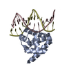


 PDBj
PDBj


 Assembly
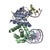
 Mass: 18.015 Da / Num. of mol.: 84 / Source method: isolated from a natural source / Formula: H2O
 Sample preparation
 / Beamline: 21-ID-D / Wavelength: 1 Å
 Processing