 Movie
Movie Controller

[English] 日本語
 Yorodumi
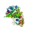
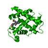
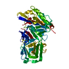
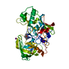
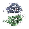
- PDB-3gd0: Crystal structure of laminaripentaose-producing beta-1,3-glucanase -
+ Open data

- Basic information
| Entry | Database: PDB / ID: 3gd0 | ||||||
|---|---|---|---|---|---|---|---|
| Title | Crystal structure of laminaripentaose-producing beta-1,3-glucanase | ||||||
 Components Components | Laminaripentaose-producing beta-1,3-guluase (LPHase) | ||||||
 Keywords Keywords | HYDROLASE / Glycoside hydrolase / Laminaripentaose-producing beta-1 / 3-glucnase / Multi-wavelength anomalous dispersion (MAD) method | ||||||
| Function / homology |  Function and homology information Function and homology informationBeta-1,3-glucanase, C-terminal domain / Beta-1,3-glucanase, N-terminal, subdomain 2 / Beta-1,3-glucanase, N-terminal / Glucan endo-1,3-beta-glucosidase / Beta-1,3-glucanase / Glycosyl hydrolases family 64 (GH64) domain profile. / Metal Transport, Frataxin; Chain A / Thaumatin / Thaumatin / Osmotin/thaumatin-like superfamily ...Beta-1,3-glucanase, C-terminal domain / Beta-1,3-glucanase, N-terminal, subdomain 2 / Beta-1,3-glucanase, N-terminal / Glucan endo-1,3-beta-glucosidase / Beta-1,3-glucanase / Glycosyl hydrolases family 64 (GH64) domain profile. / Metal Transport, Frataxin; Chain A / Thaumatin / Thaumatin / Osmotin/thaumatin-like superfamily / Sandwich / 2-Layer Sandwich / Mainly Beta / Alpha Beta Similarity search - Domain/homology | ||||||
| Biological species |  Streptomyces matensis (bacteria) Streptomyces matensis (bacteria) | ||||||
| Method |  X-RAY DIFFRACTION / SYNCHROTRON / MAD / Resolution: 1.62 Å X-RAY DIFFRACTION / SYNCHROTRON / MAD / Resolution: 1.62 Å | ||||||
 Authors Authors | Wu, H.M. / Hsu, M.T. / Liu, S.W. / Lai, C.C. / Li, Y.K. / Wang, W.C. | ||||||
 Citation Citation | Journal: J.Biol.Chem. / Year: 2009 Title: Structure, mechanistic action, and essential residues of a GH-64 enzyme, laminaripentaose-producing beta-1,3-glucanase. Authors: Wu, H.M. / Liu, S.W. / Hsu, M.T. / Hung, C.L. / Lai, C.C. / Cheng, W.C. / Wang, H.J. / Li, Y.K. / Wang, W.C. | ||||||
| History |
|
- Structure visualization
| Structure viewer | Molecule: MolmilJmol/JSmol |
|---|
- Downloads & links
-Download
| PDBx/mmCIF format | 3gd0.cif.gz | 100.2 KB | Display | PDBx/mmCIF format |
|---|---|---|---|---|
| PDB format | pdb3gd0.ent.gz | 73.5 KB | Display | PDB format |
| PDBx/mmJSON format | 3gd0.json.gz | Tree view | PDBx/mmJSON format | |
| Others |  Other downloads Other downloads |
-Validation report
| Arichive directory | https://data.pdbj.org/pub/pdb/validation_reports/gd/3gd0ftp://data.pdbj.org/pub/pdb/validation_reports/gd/3gd0 | HTTPS FTP |
|---|
-Related structure data


-Links
 PDBj
PDBj
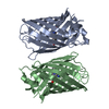
- Assembly
| Deposited unit | 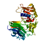
| ||||||||
|---|---|---|---|---|---|---|---|---|---|
| 1 |
| ||||||||
| Unit cell |
|
-Components
| #1: Protein | Mass: 39592.910 Da / Num. of mol.: 1 Source method: isolated from a genetically manipulated source Source: (gene. exp.) Streptomyces matensis (bacteria) / Strain: DIC-108 / Gene: LPHase / Plasmid: pREST_A / Production host: References: UniProt: Q9Z4I2, glucan endo-1,3-beta-D-glucosidase |
|---|---|
| #2: Water | ChemComp-HOH /  Mass: 18.015 Da / Num. of mol.: 730 / Source method: isolated from a natural source / Formula: H2O Mass: 18.015 Da / Num. of mol.: 730 / Source method: isolated from a natural source / Formula: H2O |
-Experimental details
-Experiment
| Experiment | Method: X-RAY DIFFRACTION / Number of used crystals: 2 |
|---|
- Sample preparation
| Crystal |
| |||||||||
|---|---|---|---|---|---|---|---|---|---|---|
| Crystal grow | Temperature: 293 K / Method: liquid diffusion / pH: 6.5 Details: 0.15M ammonium sulfate, 30% PEG MME5000, 0.1M MES (pH6.5), LIQUID DIFFUSION, temperature 293K |
-Data collection
| Diffraction |
| ||||||||||||||||||
|---|---|---|---|---|---|---|---|---|---|---|---|---|---|---|---|---|---|---|---|
| Diffraction source |
| ||||||||||||||||||
| Detector |
| ||||||||||||||||||
| Radiation |
| ||||||||||||||||||
| Radiation wavelength |
| ||||||||||||||||||
| Reflection | Resolution: 1.62→29.7 Å / Num. all: 54287 / Num. obs: 54285 / % possible obs: 100 % / Observed criterion σ(F): 28.5 / Observed criterion σ(I): 16.8 / Redundancy: 14 % / Biso Wilson estimate: 17.1 Å2 / Rmerge(I) obs: 0.06 / Rsym value: 0.047 / Net I/σ(I): 43.9 | ||||||||||||||||||
| Reflection shell | Resolution: 1.62→1.68 Å / Redundancy: 12.3 % / Rmerge(I) obs: 0.336 / Mean I/σ(I) obs: 8.4 / Num. unique all: 5354 / Rsym value: 0.3 / % possible all: 100 |


- Processing
| Software |
| |||||||||||||||||||||||||||||||||||||||||||||||||||||||||||||||||
|---|---|---|---|---|---|---|---|---|---|---|---|---|---|---|---|---|---|---|---|---|---|---|---|---|---|---|---|---|---|---|---|---|---|---|---|---|---|---|---|---|---|---|---|---|---|---|---|---|---|---|---|---|---|---|---|---|---|---|---|---|---|---|---|---|---|---|
| Refinement | Method to determine structure: MAD / Resolution: 1.62→29.7 Å / Cor.coef. Fo:Fc: 0.954 / Cor.coef. Fo:Fc free: 0.934 / SU B: 1.477 / SU ML: 0.053 / Cross valid method: THROUGHOUT / ESU R: 0.09 / ESU R Free: 0.093 / Stereochemistry target values: MAXIMUM LIKELIHOOD / Details: HYDROGENS HAVE BEEN ADDED IN THE RIDING POSITIONS
| |||||||||||||||||||||||||||||||||||||||||||||||||||||||||||||||||
| Solvent computation | Ion probe radii: 0.8 Å / Shrinkage radii: 0.8 Å / VDW probe radii: 1.2 Å / Solvent model: MASK | |||||||||||||||||||||||||||||||||||||||||||||||||||||||||||||||||
| Displacement parameters | Biso mean: 17.074 Å2
| |||||||||||||||||||||||||||||||||||||||||||||||||||||||||||||||||
| Refinement step | Cycle: LAST / Resolution: 1.62→29.7 Å
| |||||||||||||||||||||||||||||||||||||||||||||||||||||||||||||||||
| Refine LS restraints |
| |||||||||||||||||||||||||||||||||||||||||||||||||||||||||||||||||
| LS refinement shell | Resolution: 1.62→1.662 Å / Total num. of bins used: 20
|
