 Movie
Movie Controller

[English] 日本語
 Yorodumi
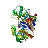
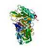
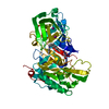
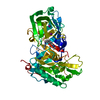
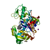
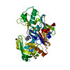
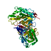
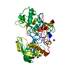
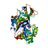
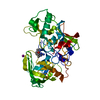
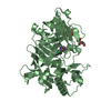
- PDB-2of0: X-ray crystal structure of beta secretase complexed with compound 5 -
+ Open data

- Basic information
| Entry | Database: PDB / ID: 2of0 | ||||||
|---|---|---|---|---|---|---|---|
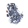
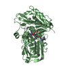
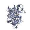
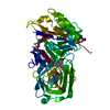
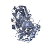
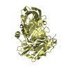
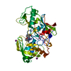
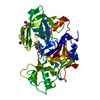
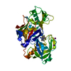
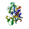
| Title | X-ray crystal structure of beta secretase complexed with compound 5 | ||||||
 Components Components | Beta-secretase 1 | ||||||
 Keywords Keywords | HYDROLASE / ALTERNATIVE SPLICING / ALZHEIMER'S DISEASE / ASPARTIC PROTEASE / ASPARTYL PROTEASE / BASE / BETA-SECRETASE / GLYCOPROTEIN / MEMAPSIN 2 / TRANSMEMBRANE / ZYMOGEN | ||||||
| Function / homology |  Function and homology information Function and homology informationmemapsin 2 / Golgi-associated vesicle lumen / beta-aspartyl-peptidase activity / signaling receptor ligand precursor processing / amyloid-beta formation / amyloid precursor protein catabolic process / membrane protein ectodomain proteolysis / amyloid-beta metabolic process / detection of mechanical stimulus involved in sensory perception of pain / response to insulin-like growth factor stimulus ...memapsin 2 / Golgi-associated vesicle lumen / beta-aspartyl-peptidase activity / signaling receptor ligand precursor processing / amyloid-beta formation / amyloid precursor protein catabolic process / membrane protein ectodomain proteolysis / amyloid-beta metabolic process / detection of mechanical stimulus involved in sensory perception of pain / response to insulin-like growth factor stimulus / prepulse inhibition / cellular response to manganese ion / multivesicular body / swimming behavior / cellular response to copper ion / presynaptic modulation of chemical synaptic transmission / hippocampal mossy fiber to CA3 synapse / protein serine/threonine kinase binding / trans-Golgi network / protein processing / recycling endosome / response to lead ion / cellular response to amyloid-beta / synaptic vesicle / late endosome / peptidase activity / positive regulation of neuron apoptotic process / amyloid-beta binding / endopeptidase activity / amyloid fibril formation / aspartic-type endopeptidase activity / early endosome / lysosome / endosome / endosome membrane / membrane raft / endoplasmic reticulum lumen / Amyloid fiber formation / axon / neuronal cell body / dendrite / enzyme binding / cell surface / Golgi apparatus / proteolysis / membrane / plasma membrane Similarity search - Function | ||||||
| Biological species |  Homo sapiens (human) Homo sapiens (human) | ||||||
| Method |  X-RAY DIFFRACTION / MOLECULAR REPLACEMENT / Resolution: 2.25 Å X-RAY DIFFRACTION / MOLECULAR REPLACEMENT / Resolution: 2.25 Å | ||||||
 Authors Authors | Patel, S. | ||||||
 Citation Citation | Journal: J.Med.Chem. / Year: 2007 Title: Application of fragment screening by X-ray crystallography to beta-Secretase. Authors: Murray, C.W. / Callaghan, O. / Chessari, G. / Cleasby, A. / Congreve, M. / Frederickson, M. / Hartshorn, M.J. / McMenamin, R. / Patel, S. / Wallis, N. | ||||||
| History |
|
- Structure visualization
| Structure viewer | Molecule: MolmilJmol/JSmol |
|---|
- Downloads & links
-Download
| PDBx/mmCIF format | 2of0.cif.gz | 95 KB | Display | PDBx/mmCIF format |
|---|---|---|---|---|
| PDB format | pdb2of0.ent.gz | 71.4 KB | Display | PDB format |
| PDBx/mmJSON format | 2of0.json.gz | Tree view | PDBx/mmJSON format | |
| Others |  Other downloads Other downloads |
-Validation report
| Arichive directory | https://data.pdbj.org/pub/pdb/validation_reports/of/2of0ftp://data.pdbj.org/pub/pdb/validation_reports/of/2of0 | HTTPS FTP |
|---|
-Related structure data
| Related structure data |  2ohkC  2ohlC  2ohmC 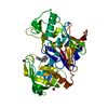 2ohnC  1w50S C: citing same article ( S: Starting model for refinement |
|---|---|
| Similar structure data |


-Links
 PDBj
PDBj

- Assembly
| Deposited unit | 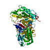
| ||||||||
|---|---|---|---|---|---|---|---|---|---|
| 1 |
| ||||||||
| Unit cell |
| ||||||||
| Details | The biological assembly is a monomer |
-Components
| #1: Protein | Mass: 44841.395 Da / Num. of mol.: 1 / Fragment: protease domain / Mutation: R56K, R57K Source method: isolated from a genetically manipulated source Source: (gene. exp.) Homo sapiens (human) / Gene: BACE1, BACE / Plasmid: pET / Species (production host): Escherichia coli / Production host:  | ||||||||
|---|---|---|---|---|---|---|---|---|---|
| #2: Chemical |   Mass: 126.904 Da / Num. of mol.: 3 / Source method: obtained synthetically / Formula: I Mass: 126.904 Da / Num. of mol.: 3 / Source method: obtained synthetically / Formula: I#3: Chemical | ChemComp-DMS / |   Mass: 78.133 Da / Num. of mol.: 1 / Source method: obtained synthetically / Formula: C2H6OS / Comment: DMSO, precipitant*YM Mass: 78.133 Da / Num. of mol.: 1 / Source method: obtained synthetically / Formula: C2H6OS / Comment: DMSO, precipitant*YM#4: Chemical | ChemComp-CMZ / ( |   Mass: 265.348 Da / Num. of mol.: 1 / Source method: obtained synthetically / Formula: C15H23NO3 Mass: 265.348 Da / Num. of mol.: 1 / Source method: obtained synthetically / Formula: C15H23NO3#5: Water | ChemComp-HOH / |  Mass: 18.015 Da / Num. of mol.: 179 / Source method: isolated from a natural source / Formula: H2O Mass: 18.015 Da / Num. of mol.: 179 / Source method: isolated from a natural source / Formula: H2OHas protein modification | Y | |
-Experimental details
-Experiment
| Experiment | Method: X-RAY DIFFRACTION / Number of used crystals: 1 |
|---|
- Sample preparation
| Crystal | Density Matthews: 2.97 Å3/Da / Density % sol: 58.53 % |
|---|---|
| Crystal grow | Temperature: 293 K / Method: vapor diffusion, hanging drop / pH: 6.6 Details: 20-22.5% (w/v) PEG 5000 monomethylether (MME), 200 mM sodium citrate (pH 6.6), 200 mM ammonium iodide, VAPOR DIFFUSION, HANGING DROP, temperature 293K |
-Data collection
| Diffraction | Mean temperature: 100 K |
|---|---|
| Diffraction source | Source: ROTATING ANODE / Type: RIGAKU RU300 / Wavelength: 1.5418 Å |
| Detector | Type: RIGAKU RAXIS IV / Detector: IMAGE PLATE / Date: Oct 24, 2002 |
| Radiation | Protocol: SINGLE WAVELENGTH / Monochromatic (M) / Laue (L): M / Scattering type: x-ray |
| Radiation wavelength | Wavelength: 1.5418 Å / Relative weight: 1 |
| Reflection | Resolution: 2.2→38.2 Å / Num. all: 26905 / Num. obs: 24904 |
- Processing
| Software |
| |||||||||||||||||||||||||||||||||||||||||||||||||||||||||||||||||||||||||||||||||||||||||||||||||||||||||||||||||||||||||||||
|---|---|---|---|---|---|---|---|---|---|---|---|---|---|---|---|---|---|---|---|---|---|---|---|---|---|---|---|---|---|---|---|---|---|---|---|---|---|---|---|---|---|---|---|---|---|---|---|---|---|---|---|---|---|---|---|---|---|---|---|---|---|---|---|---|---|---|---|---|---|---|---|---|---|---|---|---|---|---|---|---|---|---|---|---|---|---|---|---|---|---|---|---|---|---|---|---|---|---|---|---|---|---|---|---|---|---|---|---|---|---|---|---|---|---|---|---|---|---|---|---|---|---|---|---|---|---|
| Refinement | Method to determine structure: MOLECULAR REPLACEMENT Starting model: PDB entry 1W50 Resolution: 2.25→38.19 Å / Cor.coef. Fo:Fc: 0.934 / Cor.coef. Fo:Fc free: 0.89 / SU B: 6.964 / SU ML: 0.174 / Cross valid method: THROUGHOUT / σ(F): 0 / ESU R: 0.272 / ESU R Free: 0.236 / Stereochemistry target values: MAXIMUM LIKELIHOOD / Details: HYDROGENS HAVE BEEN ADDED IN THE RIDING POSITIONS
| |||||||||||||||||||||||||||||||||||||||||||||||||||||||||||||||||||||||||||||||||||||||||||||||||||||||||||||||||||||||||||||
| Solvent computation | Ion probe radii: 0.8 Å / Shrinkage radii: 0.8 Å / VDW probe radii: 1.2 Å / Solvent model: BABINET MODEL WITH MASK | |||||||||||||||||||||||||||||||||||||||||||||||||||||||||||||||||||||||||||||||||||||||||||||||||||||||||||||||||||||||||||||
| Displacement parameters | Biso mean: 36.407 Å2
| |||||||||||||||||||||||||||||||||||||||||||||||||||||||||||||||||||||||||||||||||||||||||||||||||||||||||||||||||||||||||||||
| Refinement step | Cycle: LAST / Resolution: 2.25→38.19 Å
| |||||||||||||||||||||||||||||||||||||||||||||||||||||||||||||||||||||||||||||||||||||||||||||||||||||||||||||||||||||||||||||
| Refine LS restraints |
| |||||||||||||||||||||||||||||||||||||||||||||||||||||||||||||||||||||||||||||||||||||||||||||||||||||||||||||||||||||||||||||
| LS refinement shell | Resolution: 2.25→2.308 Å / Total num. of bins used: 20
|
