Mass: 18.015 Da / Num. of mol.: 107 / Source method: isolated from a natural source / Formula: H2O
-
Experimental details
-
Experiment
Experiment
Method: X-RAY DIFFRACTION / Number of used crystals: 1
-
Sample preparation
Crystal
Density Matthews: 3.4 Å3/Da / Density % sol: 64 % Description: THE R-FACTOR CALCULATED DURING DATA REDUCTION FOR THE HIGHEST RESOLUTION SHELL DOES NOT ACCURATELY REPRESENT THE DATA. THE DATA WERE ANISOTROPIC, AND AFTER REDUCTION WERE ANISOTROPICALLY ...Description: THE R-FACTOR CALCULATED DURING DATA REDUCTION FOR THE HIGHEST RESOLUTION SHELL DOES NOT ACCURATELY REPRESENT THE DATA. THE DATA WERE ANISOTROPIC, AND AFTER REDUCTION WERE ANISOTROPICALLY TRUNCATED WITH AN ELLIPSOIDAL BOUNDARY AT I/SIGMA=2. THIS RESULTED IN A SHELL WITH A MAJOR AXIS OF 2.4 A RESOLUTION (IN THE C* DIRECTION) AND A MINOR AXIS OF 2.8 (IN THE A* AND B* DIRECTIONS). THEREFORE ABOUT 90% OF THE REFLECTIONS WHICH WERE USED TO CALCULATE AN R-FACTOR FOR THE HIGHEST RESOLUTION SHELL WERE EXCLUDED FROM REFINEMENT.
Crystal grow
pH: 6.6 / Details: pH 6.6
Crystal
*PLUS
Crystal grow
*PLUS
Temperature: 15-22 ℃ / Method: vapor diffusion, hanging drop / PH range low: 7 / PH range high: 6
Monochromator: NI FILTER / Monochromatic (M) / Laue (L): M / Scattering type: x-ray
Radiation wavelength
Wavelength: 1.5418 Å / Relative weight: 1
Reflection
Resolution: 2.4→20 Å / Num. obs: 27135 / % possible obs: 95 % / Observed criterion σ(I): -3 / Redundancy: 3.1 % / Biso Wilson estimate: 29.1 Å2 / Rmerge(I) obs: 0.087 / Rsym value: 0.087 / Net I/σ(I): 9.4
Reflection shell
Resolution: 2.4→2.58 Å / Redundancy: 1 % / Rmerge(I) obs: 1 / Mean I/σ(I) obs: 0.7 / Rsym value: 1 / % possible all: 80
-
Processing
Software
Name
Version
Classification
CNS
0.3
refinement
DENZO
datareduction
SCALEPACK
datascaling
CNS
0.3
phasing
Refinement
Method to determine structure: HEAVY ATOM ISOMORPHOUS REPLACEMENT Resolution: 2.4→10 Å / Rfactor Rfree error: 0.006 / Data cutoff high absF: 286501.92 / Isotropic thermal model: RESTRAINED / Cross valid method: THROUGHOUT / σ(F): 0
Rfactor
Num. reflection
% reflection
Selection details
Rfree
0.274
1784
9.8 %
RANDOM
Rwork
0.244
-
-
-
obs
0.244
18121
64.7 %
-
Displacement parameters
Biso mean: 35.7 Å2
Baniso -1
Baniso -2
Baniso -3
1-
0 Å2
0 Å2
0 Å2
2-
-
0 Å2
0 Å2
3-
-
-
0 Å2
Refine analyze
Free
Obs
Luzzati coordinate error
0.41 Å
0.36 Å
Luzzati d res low
-
5 Å
Luzzati sigma a
0.4 Å
0.36 Å
Refinement step
Cycle: LAST / Resolution: 2.4→10 Å
Protein
Nucleic acid
Ligand
Solvent
Total
Num. atoms
2533
0
2
107
2642
Refine LS restraints
Refine-ID
Type
Dev ideal
Dev ideal target
X-RAY DIFFRACTION
c_bond_d
0.008
X-RAY DIFFRACTION
c_bond_d_na
X-RAY DIFFRACTION
c_bond_d_prot
X-RAY DIFFRACTION
c_angle_d
X-RAY DIFFRACTION
c_angle_d_na
X-RAY DIFFRACTION
c_angle_d_prot
X-RAY DIFFRACTION
c_angle_deg
1.5
X-RAY DIFFRACTION
c_angle_deg_na
X-RAY DIFFRACTION
c_angle_deg_prot
X-RAY DIFFRACTION
c_dihedral_angle_d
21.3
X-RAY DIFFRACTION
c_dihedral_angle_d_na
X-RAY DIFFRACTION
c_dihedral_angle_d_prot
X-RAY DIFFRACTION
c_improper_angle_d
0.86
X-RAY DIFFRACTION
c_improper_angle_d_na
X-RAY DIFFRACTION
c_improper_angle_d_prot
X-RAY DIFFRACTION
c_mcbond_it
2.15
1.5
X-RAY DIFFRACTION
c_mcangle_it
3.59
2
X-RAY DIFFRACTION
c_scbond_it
3.16
2
X-RAY DIFFRACTION
c_scangle_it
4.82
2.5
LS refinement shell
Resolution: 2.4→2.55 Å / Rfactor Rfree error: 0.056 / Total num. of bins used: 6
In the structure databanks used in Yorodumi, some data are registered as the other names, "COVID-19 virus" and "2019-nCoV". Here are the details of the virus and the list of structure data.
Jan 31, 2019. EMDB accession codes are about to change! (news from PDBe EMDB page)
EMDB accession codes are about to change! (news from PDBe EMDB page)
The allocation of 4 digits for EMDB accession codes will soon come to an end. Whilst these codes will remain in use, new EMDB accession codes will include an additional digit and will expand incrementally as the available range of codes is exhausted. The current 4-digit format prefixed with “EMD-” (i.e. EMD-XXXX) will advance to a 5-digit format (i.e. EMD-XXXXX), and so on. It is currently estimated that the 4-digit codes will be depleted around Spring 2019, at which point the 5-digit format will come into force.
The EM Navigator/Yorodumi systems omit the EMD- prefix.
Related info.:Q: What is EMD? / ID/Accession-code notation in Yorodumi/EM Navigator
Yorodumi is a browser for structure data from EMDB, PDB, SASBDB, etc.
This page is also the successor to EM Navigator detail page, and also detail information page/front-end page for Omokage search.
The word "yorodu" (or yorozu) is an old Japanese word meaning "ten thousand". "mi" (miru) is to see.
Related info.:EMDB / PDB / SASBDB / Comparison of 3 databanks / Yorodumi Search / Aug 31, 2016. New EM Navigator & Yorodumi / Yorodumi Papers / Jmol/JSmol / Function and homology information / Changes in new EM Navigator and Yorodumi
 Movie
Movie Controller


 Open data
 Basic information
 Components
 Keywords
 Function and homology information
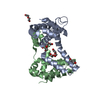
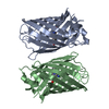
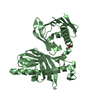
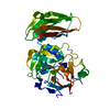
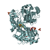
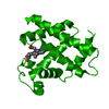
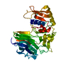
 Human poliovirus 1
 X-RAY DIFFRACTION / HEAVY ATOM ISOMORPHOUS REPLACEMENT / Resolution: 2.4 Å
  Authors
 Citation
 Structure visualization
 Downloads & links
 Other downloads
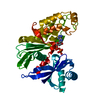


 PDBj
PDBj

 Assembly
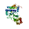


 Mass: 40.078 Da / Num. of mol.: 2 / Source method: obtained synthetically / Formula: Ca
 Mass: 18.015 Da / Num. of mol.: 107 / Source method: isolated from a natural source / Formula: H2O
 Sample preparation
 Processing