 Movie
Movie Controller

[English] 日本語
 Yorodumi
- PDB-3aar: Crystal structure of Lp1NTPDase from Legionella pneumophila in co... -
+ Open data

- Basic information
| Entry | Database: PDB / ID: 3aar | ||||||
|---|---|---|---|---|---|---|---|
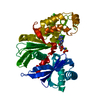
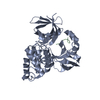
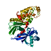
| Title | Crystal structure of Lp1NTPDase from Legionella pneumophila in complex with AMPPNP | ||||||
 Components Components | Ectonucleoside triphosphate diphosphohydrolase I | ||||||
 Keywords Keywords | HYDROLASE / adenosine triphosphatase / NTPDase | ||||||
| Function / homology |  Function and homology information Function and homology informationnucleoside diphosphate catabolic process / nucleoside diphosphate phosphatase activity / nucleotide binding / membrane / metal ion binding Similarity search - Function | ||||||
| Biological species |   Legionella pneumophila (bacteria) Legionella pneumophila (bacteria) | ||||||
| Method |  X-RAY DIFFRACTION / SYNCHROTRON / MOLECULAR REPLACEMENT / Resolution: 1.65 Å X-RAY DIFFRACTION / SYNCHROTRON / MOLECULAR REPLACEMENT / Resolution: 1.65 Å | ||||||
 Authors Authors | Ge, H. / Vivian, J.P. / Beddoe, T. / Rossjohn, J. | ||||||
 Citation Citation | Journal: Structure / Year: 2010 Title: Crystal Structure of a Legionella pneumophila Ecto -Triphosphate Diphosphohydrolase, A Structural and Functional Homolog of the Eukaryotic NTPDases Authors: Vivian, J.P. / Riedmaier, P. / Ge, H. / Le Nours, J. / Sansom, F.M. / Wilce, M.C.J. / Byres, E. / Dias, M. / Schmidberger, J.W. / Cowan, P.J. / d'Apice, A.J.F. / Hartland, E.L. / Rossjohn, J. / Beddoe, T. | ||||||
| History |
|
- Structure visualization
| Structure viewer | Molecule: MolmilJmol/JSmol |
|---|
- Downloads & links
-Download
| PDBx/mmCIF format | 3aar.cif.gz | 98.9 KB | Display | PDBx/mmCIF format |
|---|---|---|---|---|
| PDB format | pdb3aar.ent.gz | 74.3 KB | Display | PDB format |
| PDBx/mmJSON format | 3aar.json.gz | Tree view | PDBx/mmJSON format | |
| Others |  Other downloads Other downloads |
-Validation report
| Arichive directory | https://data.pdbj.org/pub/pdb/validation_reports/aa/3aarftp://data.pdbj.org/pub/pdb/validation_reports/aa/3aar | HTTPS FTP |
|---|
-Related structure data
| Related structure data |  3aapSC 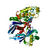 3aaqC S: Starting model for refinement C: citing same article ( |
|---|---|
| Similar structure data |


-Links
 PDBj
PDBj


- Assembly
| Deposited unit | 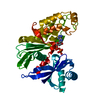
| ||||||||
|---|---|---|---|---|---|---|---|---|---|
| 1 |
| ||||||||
| Unit cell |
|
-Components
| #1: Protein | Mass: 40032.625 Da / Num. of mol.: 1 / Fragment: UNP residues 41-393 Source method: isolated from a genetically manipulated source Source: (gene. exp.) Legionella pneumophila (bacteria) / Strain: Philadelphia 1 / Gene: lpg1905 / Plasmid: pRSET / Production host: |
|---|---|
| #2: Chemical | ChemComp-ANP /   Mass: 506.196 Da / Num. of mol.: 1 / Source method: obtained synthetically / Formula: C10H17N6O12P3 / Comment: AMP-PNP, energy-carrying molecule analogue*YM Mass: 506.196 Da / Num. of mol.: 1 / Source method: obtained synthetically / Formula: C10H17N6O12P3 / Comment: AMP-PNP, energy-carrying molecule analogue*YM |
| #3: Water | ChemComp-HOH /  Mass: 18.015 Da / Num. of mol.: 494 / Source method: isolated from a natural source / Formula: H2O Mass: 18.015 Da / Num. of mol.: 494 / Source method: isolated from a natural source / Formula: H2O |
| Has protein modification | Y |
| Sequence details | ACCORDING TO AUTHOR, RESIDUE 149VAL IS A NATURAL VARIANT OF UNIPROT/TREMBL Q5ZUA2, AND WILL BE ...ACCORDING TO AUTHOR, RESIDUE 149VAL IS A NATURAL VARIANT OF UNIPROT/TREMBL Q5ZUA2, AND WILL BE REPORTED TO UNIPROT. |
-Experimental details
-Experiment
| Experiment | Method: X-RAY DIFFRACTION / Number of used crystals: 1 |
|---|
- Sample preparation
| Crystal | Density Matthews: 2.98 Å3/Da / Density % sol: 58.73 % |
|---|---|
| Crystal grow | Temperature: 294 K / Method: vapor diffusion, hanging drop / pH: 6.5 Details: 20-24%(w/v) PEG 3350, 0.2M Na Formate, 0.1M Bis-Tris propane pH 6.5, VAPOR DIFFUSION, HANGING DROP, temperature 294K |
-Data collection
| Diffraction | Mean temperature: 100 K |
|---|---|
| Diffraction source | Source: SYNCHROTRON / Site: Australian Synchrotron  / Beamline: MX1 / Wavelength: 0.954 Å / Beamline: MX1 / Wavelength: 0.954 Å |
| Detector | Type: ADSC QUANTUM 210 / Detector: CCD |
| Radiation | Protocol: SINGLE WAVELENGTH / Monochromatic (M) / Laue (L): M / Scattering type: x-ray |
| Radiation wavelength | Wavelength: 0.954 Å / Relative weight: 1 |
| Reflection | Resolution: 1.65→35 Å / Num. obs: 55849 / % possible obs: 97.6 % / Redundancy: 7.5 % / Rmerge(I) obs: 0.119 / Net I/σ(I): 14.3 |
| Reflection shell | Resolution: 1.65→1.74 Å / Redundancy: 7.3 % / Rmerge(I) obs: 0.452 / Mean I/σ(I) obs: 3.5 / % possible all: 98.8 |
- Processing
| Software |
| |||||||||||||||||||||||||||||||||||||||||||||||||||||||||||||||||||||||||||||||||||||||||||||||
|---|---|---|---|---|---|---|---|---|---|---|---|---|---|---|---|---|---|---|---|---|---|---|---|---|---|---|---|---|---|---|---|---|---|---|---|---|---|---|---|---|---|---|---|---|---|---|---|---|---|---|---|---|---|---|---|---|---|---|---|---|---|---|---|---|---|---|---|---|---|---|---|---|---|---|---|---|---|---|---|---|---|---|---|---|---|---|---|---|---|---|---|---|---|---|---|---|
| Refinement | Method to determine structure: MOLECULAR REPLACEMENT Starting model: PDB ENTRY 3AAP Resolution: 1.65→20 Å / Cor.coef. Fo:Fc: 0.959 / Cor.coef. Fo:Fc free: 0.952 / SU B: 1.63 / SU ML: 0.056 / Cross valid method: THROUGHOUT / ESU R: 0.089 / ESU R Free: 0.086 / Stereochemistry target values: MAXIMUM LIKELIHOOD / Details: HYDROGENS HAVE BEEN ADDED IN THE RIDING POSITIONS
| |||||||||||||||||||||||||||||||||||||||||||||||||||||||||||||||||||||||||||||||||||||||||||||||
| Solvent computation | Ion probe radii: 0.8 Å / Shrinkage radii: 0.8 Å / VDW probe radii: 1.2 Å / Solvent model: MASK | |||||||||||||||||||||||||||||||||||||||||||||||||||||||||||||||||||||||||||||||||||||||||||||||
| Displacement parameters | Biso mean: 17.487 Å2
| |||||||||||||||||||||||||||||||||||||||||||||||||||||||||||||||||||||||||||||||||||||||||||||||
| Refinement step | Cycle: LAST / Resolution: 1.65→20 Å
| |||||||||||||||||||||||||||||||||||||||||||||||||||||||||||||||||||||||||||||||||||||||||||||||
| Refine LS restraints |
| |||||||||||||||||||||||||||||||||||||||||||||||||||||||||||||||||||||||||||||||||||||||||||||||
| LS refinement shell | Resolution: 1.65→1.693 Å / Total num. of bins used: 20
|
