- PDB-3u7d: Crystal structure of the KRIT1/CCM1 FERM domain in complex with t... -
+
Open data
ID or keywords:
Loading...
-
Basic information
Entry
Database: PDB / ID: 3u7d
Title
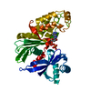
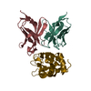
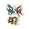
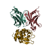
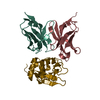
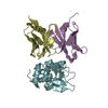
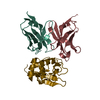
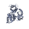
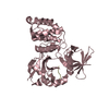
Crystal structure of the KRIT1/CCM1 FERM domain in complex with the heart of glass (HEG1) cytoplasmic tail
Components
Krev interaction trapped protein 1
Protein HEG homolog 1
Keywords
PROTEIN BINDING / PSI-Biology / Assembly / Dynamics and Evolution of Cell-Cell and Cell-Matrix Adhesions / CELLMAT / FERM domain / Rap1 effector / membrane protein cytoplasmic tail / Structural Genomics
Function / homology
Function and homology information
negative regulation of membrane permeability / : / protein localization to cell junction / GTPase regulator activity / endothelium development / integrin activation / negative regulation of endothelial cell migration / negative regulation of Rho protein signal transduction / small GTPase-mediated signal transduction / regulation of establishment of cell polarity ...negative regulation of membrane permeability / : / protein localization to cell junction / GTPase regulator activity / endothelium development / integrin activation / negative regulation of endothelial cell migration / negative regulation of Rho protein signal transduction / small GTPase-mediated signal transduction / regulation of establishment of cell polarity / negative regulation of endothelial cell proliferation / regulation of angiogenesis / negative regulation of endothelial cell apoptotic process / phosphatidylinositol-4,5-bisphosphate binding / cell redox homeostasis / negative regulation of angiogenesis / cell-cell junction / heart development / angiogenesis / microtubule binding / cytoskeleton / external side of plasma membrane / calcium ion binding / protein-containing complex / : / extracellular region / plasma membrane / cytoplasm Similarity search - Function
KRIT, N-terminal NPxY motif-rich region / Krev interaction trapped protein 1, FERM domain C-lobe / KRIT, N-terminal NPxY motif-rich domain superfamily / : / : / : / NUDIX, or N-terminal NPxY motif-rich, region of KRIT / KRIT1 ankyrin-repeats domain / KRIT1/FRMD8, FERM domain C-lobe / Acyl-CoA Binding Protein - #10 ...KRIT, N-terminal NPxY motif-rich region / Krev interaction trapped protein 1, FERM domain C-lobe / KRIT, N-terminal NPxY motif-rich domain superfamily / : / : / : / NUDIX, or N-terminal NPxY motif-rich, region of KRIT / KRIT1 ankyrin-repeats domain / KRIT1/FRMD8, FERM domain C-lobe / Acyl-CoA Binding Protein - #10 / Acyl-CoA Binding Protein / FERM central domain / : / Calcium-binding EGF domain / FERM/acyl-CoA-binding protein superfamily / Pleckstrin-homology domain (PH domain)/Phosphotyrosine-binding domain (PTB) / PH-domain like / FERM central domain / EGF-like domain / FERM superfamily, second domain / FERM domain / FERM domain profile. / Band 4.1 domain / Band 4.1 homologues / EGF-type aspartate/asparagine hydroxylation site / EGF-like calcium-binding, conserved site / Calcium-binding EGF-like domain signature. / Aspartic acid and asparagine hydroxylation site. / EGF-like calcium-binding domain / Calcium-binding EGF-like domain / Epidermal growth factor-like domain. / Phosphatidylinositol 3-kinase Catalytic Subunit; Chain A, domain 1 / EGF-like domain profile. / EGF-like domain signature 1. / EGF-like domain signature 2. / EGF-like domain / Ankyrin repeat profile. / Ankyrin repeat region circular profile. / ankyrin repeats / Ankyrin repeat / Ankyrin repeat-containing domain superfamily / Ubiquitin-like (UB roll) / PH-like domain superfamily / Roll / Roll / Up-down Bundle / Mainly Beta / Mainly Alpha / Alpha Beta Similarity search - Domain/homology
In the structure databanks used in Yorodumi, some data are registered as the other names, "COVID-19 virus" and "2019-nCoV". Here are the details of the virus and the list of structure data.
Jan 31, 2019. EMDB accession codes are about to change! (news from PDBe EMDB page)
EMDB accession codes are about to change! (news from PDBe EMDB page)
The allocation of 4 digits for EMDB accession codes will soon come to an end. Whilst these codes will remain in use, new EMDB accession codes will include an additional digit and will expand incrementally as the available range of codes is exhausted. The current 4-digit format prefixed with “EMD-” (i.e. EMD-XXXX) will advance to a 5-digit format (i.e. EMD-XXXXX), and so on. It is currently estimated that the 4-digit codes will be depleted around Spring 2019, at which point the 5-digit format will come into force.
The EM Navigator/Yorodumi systems omit the EMD- prefix.
Related info.:Q: What is EMD? / ID/Accession-code notation in Yorodumi/EM Navigator
Yorodumi is a browser for structure data from EMDB, PDB, SASBDB, etc.
This page is also the successor to EM Navigator detail page, and also detail information page/front-end page for Omokage search.
The word "yorodu" (or yorozu) is an old Japanese word meaning "ten thousand". "mi" (miru) is to see.
Related info.:EMDB / PDB / SASBDB / Comparison of 3 databanks / Yorodumi Search / Aug 31, 2016. New EM Navigator & Yorodumi / Yorodumi Papers / Jmol/JSmol / Function and homology information / Changes in new EM Navigator and Yorodumi
 Movie
Movie Controller

 Yorodumi
 Open data
 Basic information
 Components
 Keywords
 Function and homology information
 Homo sapiens (human)
 X-RAY DIFFRACTION /
  Authors
 Citation
 Structure visualization
 Downloads & links
 Other downloads
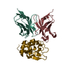


 PDBj
PDBj


 Assembly

 Mass: 18.015 Da / Num. of mol.: 10 / Source method: isolated from a natural source / Formula: H2O
 Sample preparation
 / Beamline: ID23-2 / Wavelength: 0.873 Å
 Processing