 Movie
Movie Controller


+ Open data

- Basic information
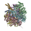
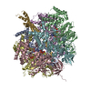
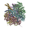
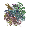
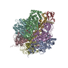
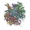
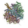
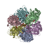
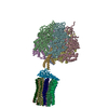
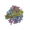
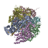
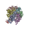
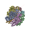
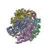
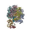
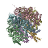
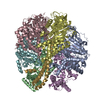
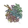
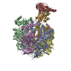
| Entry | Database: PDB / ID: 2xnd | ||||||
|---|---|---|---|---|---|---|---|
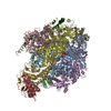
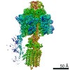
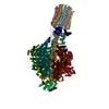
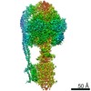
| Title | Crystal structure of bovine F1-c8 sub-complex of ATP Synthase | ||||||
 Components Components |
| ||||||
 Keywords Keywords | HYDROLASE / ATP PHOSPHORYLASE (H+ TRANSPORTING) / ATP SYNTHESIS / F1FO ATP SYNTHASE / ION TRANSPORT / P-LOOP | ||||||
| Function / homology |  Function and homology information Function and homology informationMitochondrial protein import / Formation of ATP by chemiosmotic coupling / Cristae formation / mitochondrial proton-transporting ATP synthase complex assembly / mitochondrial envelope / proton channel activity / Mitochondrial protein degradation / proton transmembrane transporter activity / proton motive force-driven ATP synthesis / proton-transporting two-sector ATPase complex, proton-transporting domain ...Mitochondrial protein import / Formation of ATP by chemiosmotic coupling / Cristae formation / mitochondrial proton-transporting ATP synthase complex assembly / mitochondrial envelope / proton channel activity / Mitochondrial protein degradation / proton transmembrane transporter activity / proton motive force-driven ATP synthesis / proton-transporting two-sector ATPase complex, proton-transporting domain / proton motive force-driven mitochondrial ATP synthesis / H+-transporting two-sector ATPase / proton-transporting ATP synthase complex / proton-transporting ATP synthase activity, rotational mechanism / proton transmembrane transport / aerobic respiration / ADP binding / mitochondrial membrane / mitochondrial inner membrane / lipid binding / structural molecule activity / ATP hydrolysis activity / mitochondrion / ATP binding / metal ion binding / plasma membrane Similarity search - Function | ||||||
| Biological species |  | ||||||
| Method |  X-RAY DIFFRACTION / SYNCHROTRON / MOLECULAR REPLACEMENT / Resolution: 3.5 Å X-RAY DIFFRACTION / SYNCHROTRON / MOLECULAR REPLACEMENT / Resolution: 3.5 Å | ||||||
 Authors Authors | Watt, I.N. / Montgomery, M.G. / Runswick, M.J. / Leslie, A.G.W. / Walker, J.E. | ||||||
 Citation Citation | Journal: Proc.Natl.Acad.Sci.USA / Year: 2010 Title: Bioenergetic Cost of Making an Adenosine Triphosphate Molecule in Animal Mitochondria. Authors: Watt, I.N. / Montgomery, M.G. / Runswick, M.J. / Leslie, A.G.W. / Walker, J.E. | ||||||
| History |
| ||||||
| Remark 700 | SHEET DETERMINATION METHOD: DSSP THE SHEETS PRESENTED AS "AA" IN EACH CHAIN ON SHEET RECORDS BELOW ... SHEET DETERMINATION METHOD: DSSP THE SHEETS PRESENTED AS "AA" IN EACH CHAIN ON SHEET RECORDS BELOW IS ACTUALLY AN 10-STRANDED BARREL THIS IS REPRESENTED BY A 11-STRANDED SHEET IN WHICH THE FIRST AND LAST STRANDS ARE IDENTICAL. THE SHEETS PRESENTED AS "DA" IN EACH CHAIN ON SHEET RECORDS BELOW IS ACTUALLY AN 6-STRANDED BARREL THIS IS REPRESENTED BY A 7-STRANDED SHEET IN WHICH THE FIRST AND LAST STRANDS ARE IDENTICAL. THE SHEET STRUCTURE OF THIS MOLECULE IS BIFURCATED. IN ORDER TO REPRESENT THIS FEATURE IN THE SHEET RECORDS BELOW, TWO SHEETS ARE DEFINED. |
- Structure visualization
| Structure viewer | Molecule: MolmilJmol/JSmol |
|---|
- Downloads & links
-Download
| PDBx/mmCIF format | 2xnd.cif.gz | 689.5 KB | Display | PDBx/mmCIF format |
|---|---|---|---|---|
| PDB format | pdb2xnd.ent.gz | 556.2 KB | Display | PDB format |
| PDBx/mmJSON format | 2xnd.json.gz | Tree view | PDBx/mmJSON format | |
| Others |  Other downloads Other downloads |
-Validation report
| Arichive directory | https://data.pdbj.org/pub/pdb/validation_reports/xn/2xndftp://data.pdbj.org/pub/pdb/validation_reports/xn/2xnd | HTTPS FTP |
|---|
-Related structure data
| Related structure data | 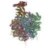 1e79S S: Starting model for refinement |
|---|---|
| Similar structure data |


-Links
 PDBj
PDBj


- Assembly
| Deposited unit | 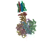
| ||||||||
|---|---|---|---|---|---|---|---|---|---|
| 1 |
| ||||||||
| Unit cell |
|
-Components
-ATP SYNTHASE SUBUNIT ... , 5 types, 9 molecules ABCDEFGHI
| #1: Protein | Mass: 53386.066 Da / Num. of mol.: 3 / Fragment: RESIDUES 62-553 / Source method: isolated from a natural source / Source: (natural) References: UniProt: P19483, H+-transporting two-sector ATPase #2: Protein | Mass: 50365.371 Da / Num. of mol.: 3 / Fragment: RESIDUES 59-525 / Source method: isolated from a natural source / Source: (natural) References: UniProt: P00829, H+-transporting two-sector ATPase #3: Protein | | Mass: 30185.674 Da / Num. of mol.: 1 / Fragment: RESIDUES 26-297 / Source method: isolated from a natural source / Source: (natural) References: UniProt: P05631, H+-transporting two-sector ATPase #4: Protein | | Mass: 13811.496 Da / Num. of mol.: 1 / Fragment: RESIDUES 37-167 / Source method: isolated from a natural source / Source: (natural) References: UniProt: P05630, H+-transporting two-sector ATPase #5: Protein/peptide | | Mass: 5275.220 Da / Num. of mol.: 1 / Fragment: RESIDUES 2-48 / Source method: isolated from a natural source / Source: (natural) References: UniProt: P05632, H+-transporting two-sector ATPase |
|---|
-Protein , 1 types, 8 molecules JKLMNOPQ
| #6: Protein | Mass: 7293.593 Da / Num. of mol.: 8 / Fragment: RESIDUES 63-134 / Source method: isolated from a natural source / Source: (natural) References: UniProt: P32876, H+-transporting two-sector ATPase |
|---|
-Non-polymers , 4 types, 12 molecules 


| #7: Chemical | ChemComp-ANP /  Mass: 506.196 Da / Num. of mol.: 5 / Source method: obtained synthetically / Formula: C10H17N6O12P3 / Comment: AMP-PNP, energy-carrying molecule analogue*YM Mass: 506.196 Da / Num. of mol.: 5 / Source method: obtained synthetically / Formula: C10H17N6O12P3 / Comment: AMP-PNP, energy-carrying molecule analogue*YM#8: Chemical | ChemComp-MG /  Mass: 24.305 Da / Num. of mol.: 5 / Source method: obtained synthetically / Formula: Mg Mass: 24.305 Da / Num. of mol.: 5 / Source method: obtained synthetically / Formula: Mg#9: Chemical | ChemComp-GOL / |  Mass: 92.094 Da / Num. of mol.: 1 / Source method: obtained synthetically / Formula: C3H8O3 Mass: 92.094 Da / Num. of mol.: 1 / Source method: obtained synthetically / Formula: C3H8O3#10: Chemical | ChemComp-SO4 / |  Mass: 96.063 Da / Num. of mol.: 1 / Source method: obtained synthetically / Formula: SO4 Mass: 96.063 Da / Num. of mol.: 1 / Source method: obtained synthetically / Formula: SO4 |
|---|
-Details
| Sequence details | DIFFERENT RESIDUE: GLY A 481, GLY B 481, GLY C 481 THIS RESIDUE WAS IDENTIFIED AS A GLY FROM THE ...DIFFERENT RESIDUE: GLY A 481, GLY B 481, GLY C 481 THIS RESIDUE WAS IDENTIFIED |
|---|
-Experimental details
-Experiment
| Experiment | Method: X-RAY DIFFRACTION / Number of used crystals: 1 |
|---|
- Sample preparation
| Crystal | Density Matthews: 3.17 Å3/Da / Density % sol: 61.16 % / Description: NONE |
|---|---|
| Crystal grow | pH: 7 Details: CRYSTALS WERE GROWN UNDER OIL BY MIXING EQUAL VOLUMES OF PROTEIN (10MG/ML IN 20MM TRIS PH 8.0, 10% GLYCEROL, 1MM ADP, 1MM AMP-PNP, 2MM MGSO4, 0.02% NAN3, 5.7MM TDM) AND PRECIPITANT SOLUTION ...Details: CRYSTALS WERE GROWN UNDER OIL BY MIXING EQUAL VOLUMES OF PROTEIN (10MG/ML IN 20MM TRIS PH 8.0, 10% GLYCEROL, 1MM ADP, 1MM AMP-PNP, 2MM MGSO4, 0.02% NAN3, 5.7MM TDM) AND PRECIPITANT SOLUTION (50MM HEPES PH 7.0, 14% PEG4600, 50MM K2HPO4) |
-Data collection
| Diffraction | Mean temperature: 100 K |
|---|---|
| Diffraction source | Source: SYNCHROTRON / Site: SLS  / Beamline: X06SA / Wavelength: 1.0007 / Beamline: X06SA / Wavelength: 1.0007 |
| Detector | Type: MARRESEARCH / Detector: CCD / Date: May 2, 2010 |
| Radiation | Protocol: SINGLE WAVELENGTH / Monochromatic (M) / Laue (L): M / Scattering type: x-ray |
| Radiation wavelength | Wavelength: 1.0007 Å / Relative weight: 1 |
| Reflection | Resolution: 3.5→132.6 Å / Num. obs: 72959 / % possible obs: 99.6 % / Observed criterion σ(I): 0 / Redundancy: 3.6 % / Biso Wilson estimate: 71.804 Å2 / Rmerge(I) obs: 0.26 / Net I/σ(I): 3.7 |
| Reflection shell | Resolution: 3.5→3.59 Å / Redundancy: 3.5 % / Rmerge(I) obs: 0.82 / Mean I/σ(I) obs: 1.6 / % possible all: 99.5 |
- Processing
| Software |
| ||||||||||||||||||||||||||||||||||||||||||||||||||||||||||||||||||||||||||||||||||||||||||||||||||||||||||||||||||||||||||||||||||||||||||||||||||||||||||||||||||||||||||||||||||||||
|---|---|---|---|---|---|---|---|---|---|---|---|---|---|---|---|---|---|---|---|---|---|---|---|---|---|---|---|---|---|---|---|---|---|---|---|---|---|---|---|---|---|---|---|---|---|---|---|---|---|---|---|---|---|---|---|---|---|---|---|---|---|---|---|---|---|---|---|---|---|---|---|---|---|---|---|---|---|---|---|---|---|---|---|---|---|---|---|---|---|---|---|---|---|---|---|---|---|---|---|---|---|---|---|---|---|---|---|---|---|---|---|---|---|---|---|---|---|---|---|---|---|---|---|---|---|---|---|---|---|---|---|---|---|---|---|---|---|---|---|---|---|---|---|---|---|---|---|---|---|---|---|---|---|---|---|---|---|---|---|---|---|---|---|---|---|---|---|---|---|---|---|---|---|---|---|---|---|---|---|---|---|---|---|
| Refinement | Method to determine structure: MOLECULAR REPLACEMENT Starting model: PDB ENTRY 1E79 Resolution: 3.5→132.6 Å / Cor.coef. Fo:Fc: 0.851 / Cor.coef. Fo:Fc free: 0.819 / SU B: 39.542 / SU ML: 0.602 / Cross valid method: THROUGHOUT / ESU R: 0 / ESU R Free: 0.674 / Stereochemistry target values: MAXIMUM LIKELIHOOD Details: HYDROGENS HAVE BEEN ADDED IN THE RIDING POSITIONS. U VALUES REFINED INDIVIDUALLY.
| ||||||||||||||||||||||||||||||||||||||||||||||||||||||||||||||||||||||||||||||||||||||||||||||||||||||||||||||||||||||||||||||||||||||||||||||||||||||||||||||||||||||||||||||||||||||
| Solvent computation | Ion probe radii: 0.8 Å / Shrinkage radii: 0.8 Å / VDW probe radii: 1.4 Å / Solvent model: MASK | ||||||||||||||||||||||||||||||||||||||||||||||||||||||||||||||||||||||||||||||||||||||||||||||||||||||||||||||||||||||||||||||||||||||||||||||||||||||||||||||||||||||||||||||||||||||
| Displacement parameters | Biso mean: 80.15 Å2
| ||||||||||||||||||||||||||||||||||||||||||||||||||||||||||||||||||||||||||||||||||||||||||||||||||||||||||||||||||||||||||||||||||||||||||||||||||||||||||||||||||||||||||||||||||||||
| Refinement step | Cycle: LAST / Resolution: 3.5→132.6 Å
| ||||||||||||||||||||||||||||||||||||||||||||||||||||||||||||||||||||||||||||||||||||||||||||||||||||||||||||||||||||||||||||||||||||||||||||||||||||||||||||||||||||||||||||||||||||||
| Refine LS restraints |
|