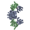
SHEET DETERMINATION METHOD: DSSP THE SHEETS PRESENTED AS "AB" IN EACH CHAIN ON SHEET RECORDS BELOW ... SHEET DETERMINATION METHOD: DSSP THE SHEETS PRESENTED AS "AB" IN EACH CHAIN ON SHEET RECORDS BELOW IS ACTUALLY AN 8-STRANDED BARREL THIS IS REPRESENTED BY A 9-STRANDED SHEET IN WHICH THE FIRST AND LAST STRANDS ARE IDENTICAL. THE SHEETS PRESENTED AS "BB" IN EACH CHAIN ON SHEET RECORDS BELOW IS ACTUALLY AN 8-STRANDED BARREL THIS IS REPRESENTED BY A 9-STRANDED SHEET IN WHICH THE FIRST AND LAST STRANDS ARE IDENTICAL.
Resolution: 2→47.5 Å / Cor.coef. Fo:Fc: 0.955 / Cor.coef. Fo:Fc free: 0.942 / SU B: 10.705 / SU ML: 0.131 / Cross valid method: THROUGHOUT / ESU R: 0.17 / ESU R Free: 0.155 / Stereochemistry target values: MAXIMUM LIKELIHOOD Details: HYDROGENS HAVE BEEN ADDED IN THE RIDING POSITIONS. U VALUES WITH TLS ADDED
Rfactor
Num. reflection
% reflection
Selection details
Rfree
0.23914
5654
4.9 %
RANDOM
Rwork
0.20288
-
-
-
obs
0.2047
108834
97.51 %
-
Solvent computation
Ion probe radii: 0.8 Å / Shrinkage radii: 0.8 Å / VDW probe radii: 1.4 Å / Solvent model: MASK
 Movie
Movie Controller


 Open data
 Basic information
 Components
 Keywords
 Function and homology information
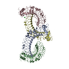
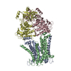
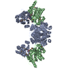
 BACTEROIDES THETAIOTAOMICRON VPI-5482 (bacteria)
 X-RAY DIFFRACTION /
  Authors
 Citation
 Structure visualization
 Downloads & links
 Other downloads


 PDBj
PDBj
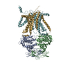
 Assembly
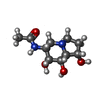
 Mass: 230.261 Da / Num. of mol.: 2 / Source method: obtained synthetically / Formula: C10H18N2O4
 Mass: 40.078 Da / Num. of mol.: 2 / Source method: obtained synthetically / Formula: Ca
 Mass: 18.015 Da / Num. of mol.: 392 / Source method: isolated from a natural source / Formula: H2O
 Sample preparation
 / Beamline: I04 / Wavelength: 0.9802
  Processing