[English] 日本語
Yorodumi
- PDB-2cho: Bacteroides thetaiotaomicron hexosaminidase with O-GlcNAcase activity -

+
Open data


ID or keywords:

Loading...

-
Basic information

Entry
Database: PDB / ID: 2cho
TitleBacteroides thetaiotaomicron hexosaminidase with O-GlcNAcase activity
Components(GLUCOSAMINIDASE) x 2
KeywordsHYDROLASE / O-GLCNACASE / N-ACETYLGLUCOSAMINE
Function / homology
Function and homology information


protein O-GlcNAcase / : / : / [protein]-3-O-(N-acetyl-D-glucosaminyl)-L-serine/L-threonine O-N-acetyl-alpha-D-glucosaminase activity / protein deglycosylation / beta-N-acetylglucosaminidase activity / carbohydrate metabolic process / identical protein binding
Similarity search - Function
: / : / Carbohydrate binding module family 32 / Bacterial GH84, post-catalytic domain / Hyaluronidase post-catalytic domain-like / Glycosyl hydrolases family 84 (GH84) domain profile. / Beta-N-acetylglucosaminidase / beta-N-acetylglucosaminidase / Chitobiase/beta-hexosaminidase domain 2-like / Chitobiase; domain 2 ...: / : / Carbohydrate binding module family 32 / Bacterial GH84, post-catalytic domain / Hyaluronidase post-catalytic domain-like / Glycosyl hydrolases family 84 (GH84) domain profile. / Beta-N-acetylglucosaminidase / beta-N-acetylglucosaminidase / Chitobiase/beta-hexosaminidase domain 2-like / Chitobiase; domain 2 / Beta-hexosaminidase, bacterial type, N-terminal / Glycosyl hydrolase family 20, domain 2 / Beta-hexosaminidase-like, domain 2 / Glycosyl hydrolase, all-beta / Methane Monooxygenase Hydroxylase; Chain G, domain 1 / Glycosidases / Glycoside hydrolase superfamily / TIM Barrel / Alpha-Beta Barrel / Up-down Bundle / 2-Layer Sandwich / Mainly Alpha / Alpha Beta
Similarity search - Domain/homology
ACETATE ION / O-GlcNAcase BT_4395
Similarity search - Component
Biological speciesBACTEROIDES THETAIOTAOMICRON (bacteria)
MethodX-RAY DIFFRACTION / SYNCHROTRON / OTHER / Resolution: 1.85 Å
AuthorsDennis, R.J. / Taylor, E.J. / Macauley, M.S. / Stubbs, K.A. / Turkenburg, J.P. / Hart, S.J. / Black, G.N. / Vocadlo, D.J. / Davies, G.J.
CitationJournal: Nat.Struct.Mol.Biol. / Year: 2006
Title: Structure and Mechanism of a Bacterial B-Glucosaminidase Having O-Glcnacase Activity
Authors: Dennis, R.J. / Taylor, E.J. / Macauley, M.S. / Stubbs, K.A. / Turkenburg, J.P. / Hart, S.J. / Black, G.N. / Vocadlo, D.J. / Davies, G.J.
History
DepositionMar 16, 2006Deposition site: PDBE / Processing site: PDBE
Revision 1.0Jun 19, 2006Provider: repository / Type: Initial release
Revision 1.1May 8, 2011Group: Version format compliance
Revision 1.2Jul 13, 2011Group: Version format compliance
Revision 1.3May 8, 2024Group: Data collection / Database references ...Data collection / Database references / Derived calculations / Other
Category: chem_comp_atom / chem_comp_bond ...chem_comp_atom / chem_comp_bond / database_2 / pdbx_database_status / pdbx_struct_conn_angle / struct_conn
Item: _database_2.pdbx_DOI / _database_2.pdbx_database_accession ..._database_2.pdbx_DOI / _database_2.pdbx_database_accession / _pdbx_database_status.status_code_sf / _pdbx_struct_conn_angle.ptnr1_auth_comp_id / _pdbx_struct_conn_angle.ptnr1_auth_seq_id / _pdbx_struct_conn_angle.ptnr1_label_asym_id / _pdbx_struct_conn_angle.ptnr1_label_atom_id / _pdbx_struct_conn_angle.ptnr1_label_comp_id / _pdbx_struct_conn_angle.ptnr1_label_seq_id / _pdbx_struct_conn_angle.ptnr3_auth_comp_id / _pdbx_struct_conn_angle.ptnr3_auth_seq_id / _pdbx_struct_conn_angle.ptnr3_label_asym_id / _pdbx_struct_conn_angle.ptnr3_label_atom_id / _pdbx_struct_conn_angle.ptnr3_label_comp_id / _pdbx_struct_conn_angle.ptnr3_label_seq_id / _pdbx_struct_conn_angle.value / _struct_conn.pdbx_dist_value / _struct_conn.ptnr1_auth_comp_id / _struct_conn.ptnr1_auth_seq_id / _struct_conn.ptnr1_label_asym_id / _struct_conn.ptnr1_label_atom_id / _struct_conn.ptnr1_label_comp_id / _struct_conn.ptnr1_label_seq_id / _struct_conn.ptnr2_auth_comp_id / _struct_conn.ptnr2_auth_seq_id / _struct_conn.ptnr2_label_asym_id / _struct_conn.ptnr2_label_atom_id / _struct_conn.ptnr2_label_comp_id / _struct_conn.ptnr2_label_seq_id
Remark 700 
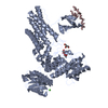
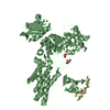
SHEET DETERMINATION METHOD: DSSP THE SHEETS PRESENTED AS "AB" IN EACH CHAIN ON SHEET RECORDS BELOW ... SHEET DETERMINATION METHOD: DSSP THE SHEETS PRESENTED AS "AB" IN EACH CHAIN ON SHEET RECORDS BELOW IS ACTUALLY AN 8-STRANDED BARREL THIS IS REPRESENTED BY A 9-STRANDED SHEET IN WHICH THE FIRST AND LAST STRANDS ARE IDENTICAL. THE SHEETS PRESENTED AS "BB" IN EACH CHAIN ON SHEET RECORDS BELOW IS ACTUALLY AN 8-STRANDED BARREL THIS IS REPRESENTED BY A 9-STRANDED SHEET IN WHICH THE FIRST AND LAST STRANDS ARE IDENTICAL. THE SHEET STRUCTURE OF THIS MOLECULE IS BIFURCATED. IN ORDER TO REPRESENT THIS FEATURE IN THE SHEET RECORDS BELOW, TWO SHEETS ARE DEFINED.

-
Structure visualization

Structure viewerMolecule:
MolmilJmol/JSmol

Downloads & links

-
Assembly

Deposited unit
A: GLUCOSAMINIDASE
B: GLUCOSAMINIDASE
C: GLUCOSAMINIDASE
D: GLUCOSAMINIDASE
hetero molecules


Theoretical massNumber of molelcules
Total (without water)167,47612
Polymers166,9094
Non-polymers5678
Water21,4201189
1
A: GLUCOSAMINIDASE
C: GLUCOSAMINIDASE
hetero molecules


Theoretical massNumber of molelcules
Total (without water)83,7386
Polymers83,4552
Non-polymers2834
Water362
TypeNameSymmetry operationNumber
identity operation1_555x,y,z1
MethodPQS
2
B: GLUCOSAMINIDASE
D: GLUCOSAMINIDASE
hetero molecules


Theoretical massNumber of molelcules
Total (without water)83,7386
Polymers83,4552
Non-polymers2834
Water362
TypeNameSymmetry operationNumber
identity operation1_555x,y,z1
MethodPQS
Unit cell
Length a, b, c (Å)51.685, 93.454, 98.828
Angle α, β, γ (deg.)75.51, 94.15, 77.15
Int Tables number1
Space group name H-MP1
Noncrystallographic symmetry (NCS)NCS domain:
IDEns-IDDetails
11A
21B

NCS domain segments:
Dom-IDComponent-IDEns-IDRefine codeAuth asym-IDAuth seq-ID
1114A1 - 999
2114B1 - 999
DetailsCHAINS C AND D ARE 2 CHAINS OF UNKNOWN ATOMS AND ARE PARTOF CHAINS A AND B, THEREFORE CHAIN A AND C IS A MONOMERAND NOT A DIMER

-
Components

-
Protein / Protein/peptide , 2 types, 4 molecules ABCD

#1: Protein GLUCOSAMINIDASE / HEXOSAMINIASE


Mass: 82330.133 Da / Num. of mol.: 2 / Fragment: RESIDUES 22-737
Source method: isolated from a genetically manipulated source
Source: (gene. exp.) BACTEROIDES THETAIOTAOMICRON (bacteria)
Strain: VPI-5482 / Plasmid: PET28A / Production host: ESCHERICHIA COLI (E. coli) / Strain (production host): BL21 / References: UniProt: Q89ZI2, beta-N-acetylhexosaminidase
#2: Protein/peptide GLUCOSAMINIDASE / HEXOSAMINIASE


Mass: 1124.378 Da / Num. of mol.: 2 / Fragment: C TERMINUS, RESIDUES 650-653,660-669
Source method: isolated from a genetically manipulated source
Source: (gene. exp.) BACTEROIDES THETAIOTAOMICRON (bacteria)
Strain: VPI-5482 / Plasmid: PET28A / Production host: ESCHERICHIA COLI (E. coli) / Strain (production host): BL21 / References: beta-N-acetylhexosaminidase

-
Non-polymers , 4 types, 1197 molecules

#3: Chemical ChemComp-CA / CALCIUM ION


Mass: 40.078 Da / Num. of mol.: 2 / Source method: obtained synthetically / Formula: Ca
#4: Chemical ChemComp-ACT / ACETATE ION


Mass: 59.044 Da / Num. of mol.: 2 / Source method: obtained synthetically / Formula: C2H3O2
#5: Chemical
ChemComp-GOL / GLYCEROL / GLYCERIN / PROPANE-1,2,3-TRIOL


Mass: 92.094 Da / Num. of mol.: 4 / Source method: obtained synthetically / Formula: C3H8O3
#6: Water ChemComp-HOH / water


Mass: 18.015 Da / Num. of mol.: 1189 / Source method: isolated from a natural source / Formula: H2O

-
Details

Sequence detailsRESIDUES IN DISORDERED C TERMINUS MODELLED AS POLYALANINE CHAINS C AND D ARE THE UNKNOWN RESIDUES ...RESIDUES IN DISORDERED C TERMINUS MODELLED AS POLYALANINE CHAINS C AND D ARE THE UNKNOWN RESIDUES OF THE C TERMINUS OF CHAINS A AND B

-
Experimental details

-
Experiment

ExperimentMethod: X-RAY DIFFRACTION / Number of used crystals: 1

-
Sample preparation

CrystalDensity Matthews: 2.7 Å3/Da / Density % sol: 54.13 %
Crystal growpH: 6
Details: 0.5M NA ACETATE, 15% PEG 3500, 0.1M MES, PH6.0 20% GLYCEROL, pH 6.00

-
Data collection

DiffractionMean temperature: 100 K
Diffraction sourceSource: SYNCHROTRON / Site: ESRF / Beamline: ID14-4 / Wavelength: 0.9393
DetectorType: ADSC CCD / Detector: CCD
RadiationProtocol: SINGLE WAVELENGTH / Monochromatic (M) / Laue (L): M / Scattering type: x-ray
Radiation wavelengthWavelength: 0.9393 Å / Relative weight: 1
ReflectionResolution: 1.85→30 Å / Num. obs: 135175 / % possible obs: 97 % / Observed criterion σ(I): 0 / Redundancy: 3.9 % / Rmerge(I) obs: 0.09 / Net I/σ(I): 13
Reflection shellResolution: 1.85→1.95 Å / Redundancy: 3.8 % / Rmerge(I) obs: 0.49 / Mean I/σ(I) obs: 3 / % possible all: 96

-
Processing

Software
NameVersionClassification
REFMAC5.2.0005refinement
MOSFLMdata reduction
SCALAdata scaling
RefinementMethod to determine structure: OTHER / Resolution: 1.85→37.74 Å / Cor.coef. Fo:Fc: 0.951 / Cor.coef. Fo:Fc free: 0.927 / SU B: 2.913 / SU ML: 0.088 / Cross valid method: THROUGHOUT / ESU R: 0.125 / ESU R Free: 0.124 / Stereochemistry target values: MAXIMUM LIKELIHOOD
Details: HYDROGENS HAVE BEEN ADDED IN THE RIDING POSITIONS. FOLLOWING POST PROOF-SETTING REVISIONS INFORMED BY THE ANALYSIS PROVIDED BY THE PDB, THE STATISTICS OF THE ACTUALLY COORDINATE FILE ARE ...Details: HYDROGENS HAVE BEEN ADDED IN THE RIDING POSITIONS. FOLLOWING POST PROOF-SETTING REVISIONS INFORMED BY THE ANALYSIS PROVIDED BY THE PDB, THE STATISTICS OF THE ACTUALLY COORDINATE FILE ARE SLIGHTLY DIFFERENT TO THAT REPORTED IN THE PUBLICATION.
RfactorNum. reflection% reflectionSelection details
Rfree0.22 7154 5 %RANDOM
Rwork0.18 ---
obs0.182 135175 96.4 %-
Solvent computationIon probe radii: 0.8 Å / Shrinkage radii: 0.8 Å / VDW probe radii: 1.2 Å / Solvent model: MASK
Displacement parametersBiso mean: 22.93 Å2
Baniso -1Baniso -2Baniso -3
1-0 Å2-0.02 Å2-0.01 Å2
2---0.01 Å2-0.05 Å2
3---0.04 Å2
Refinement stepCycle: LAST / Resolution: 1.85→37.74 Å
ProteinNucleic acidLigandSolventTotal
Num. atoms10536 0 34 1189 11759
Refine LS restraints
Refine-IDTypeDev idealDev ideal targetNumber
X-RAY DIFFRACTIONr_bond_refined_d0.0140.02210871
X-RAY DIFFRACTIONr_bond_other_d0.0050.029646
X-RAY DIFFRACTIONr_angle_refined_deg1.4331.95414742
X-RAY DIFFRACTIONr_angle_other_deg0.829322569
X-RAY DIFFRACTIONr_dihedral_angle_1_deg7.84251310
X-RAY DIFFRACTIONr_dihedral_angle_2_deg36.40324.878531
X-RAY DIFFRACTIONr_dihedral_angle_3_deg14.548151863
X-RAY DIFFRACTIONr_dihedral_angle_4_deg15.9661550
X-RAY DIFFRACTIONr_chiral_restr0.0880.21573
X-RAY DIFFRACTIONr_gen_planes_refined0.0060.0212020
X-RAY DIFFRACTIONr_gen_planes_other0.0010.022144
X-RAY DIFFRACTIONr_nbd_refined0.2120.22239
X-RAY DIFFRACTIONr_nbd_other0.190.29831
X-RAY DIFFRACTIONr_nbtor_refined0.1790.25326
X-RAY DIFFRACTIONr_nbtor_other0.0890.25517
X-RAY DIFFRACTIONr_xyhbond_nbd_refined0.190.2978
X-RAY DIFFRACTIONr_xyhbond_nbd_other
X-RAY DIFFRACTIONr_metal_ion_refined0.0840.24
X-RAY DIFFRACTIONr_metal_ion_other
X-RAY DIFFRACTIONr_symmetry_vdw_refined0.1810.223
X-RAY DIFFRACTIONr_symmetry_vdw_other0.2410.265
X-RAY DIFFRACTIONr_symmetry_hbond_refined0.2430.224
X-RAY DIFFRACTIONr_symmetry_hbond_other
X-RAY DIFFRACTIONr_symmetry_metal_ion_refined
X-RAY DIFFRACTIONr_symmetry_metal_ion_other
X-RAY DIFFRACTIONr_mcbond_it1.2411.56802
X-RAY DIFFRACTIONr_mcbond_other0.2211.52620
X-RAY DIFFRACTIONr_mcangle_it1.509210609
X-RAY DIFFRACTIONr_mcangle_other
X-RAY DIFFRACTIONr_scbond_it2.54134789
X-RAY DIFFRACTIONr_scbond_other
X-RAY DIFFRACTIONr_scangle_it3.7754.54127
X-RAY DIFFRACTIONr_scangle_other
X-RAY DIFFRACTIONr_long_range_B_refined
X-RAY DIFFRACTIONr_long_range_B_other
X-RAY DIFFRACTIONr_rigid_bond_restr
X-RAY DIFFRACTIONr_sphericity_free
X-RAY DIFFRACTIONr_sphericity_bonded
Refine LS restraints NCS

Dom-ID: 1 / Auth asym-ID: A / Ens-ID: 1 / Number: 10035 / Refine-ID: X-RAY DIFFRACTION

TypeRms dev position (Å)Weight position
medium positional0.320.5
medium thermal0.632
LS refinement shellResolution: 1.85→1.9 Å / Total num. of bins used: 20
RfactorNum. reflection% reflection
Rfree0.289 472 -
Rwork0.25 9982 -
obs--96.02 %

+
About Yorodumi

-
News

-
Feb 9, 2022. New format data for meta-information of EMDB entries

New format data for meta-information of EMDB entries

  • Version 3 of the EMDB header file is now the official format.
  • The previous official version 1.9 will be removed from the archive.

Related info.:EMDB header

External links:wwPDB to switch to version 3 of the EMDB data model

-
Aug 12, 2020. Covid-19 info

Covid-19 info

URL: https://pdbjlvh1.pdbj.org/emnavi/covid19.php

New page: Covid-19 featured information page in EM Navigator.

Related info.:Covid-19 info / Mar 5, 2020. Novel coronavirus structure data

+
Mar 5, 2020. Novel coronavirus structure data

Novel coronavirus structure data

Related info.:Yorodumi Speices / Aug 12, 2020. Covid-19 info

External links:COVID-19 featured content - PDBj / Molecule of the Month (242):Coronavirus Proteases

+
Jan 31, 2019. EMDB accession codes are about to change! (news from PDBe EMDB page)

EMDB accession codes are about to change! (news from PDBe EMDB page)

  • The allocation of 4 digits for EMDB accession codes will soon come to an end. Whilst these codes will remain in use, new EMDB accession codes will include an additional digit and will expand incrementally as the available range of codes is exhausted. The current 4-digit format prefixed with “EMD-” (i.e. EMD-XXXX) will advance to a 5-digit format (i.e. EMD-XXXXX), and so on. It is currently estimated that the 4-digit codes will be depleted around Spring 2019, at which point the 5-digit format will come into force.
  • The EM Navigator/Yorodumi systems omit the EMD- prefix.

Related info.:Q: What is EMD? / ID/Accession-code notation in Yorodumi/EM Navigator

External links:EMDB Accession Codes are Changing Soon! / Contact to PDBj

+
Jul 12, 2017. Major update of PDB

Major update of PDB

  • wwPDB released updated PDB data conforming to the new PDBx/mmCIF dictionary.
  • This is a major update changing the version number from 4 to 5, and with Remediation, in which all the entries are updated.
  • In this update, many items about electron microscopy experimental information are reorganized (e.g. em_software).
  • Now, EM Navigator and Yorodumi are based on the updated data.

External links:wwPDB Remediation / Enriched Model Files Conforming to OneDep Data Standards Now Available in the PDB FTP Archive

-
Yorodumi

Thousand views of thousand structures

  • Yorodumi is a browser for structure data from EMDB, PDB, SASBDB, etc.
  • This page is also the successor to EM Navigator detail page, and also detail information page/front-end page for Omokage search.
  • The word "yorodu" (or yorozu) is an old Japanese word meaning "ten thousand". "mi" (miru) is to see.

Related info.:EMDB / PDB / SASBDB / Comparison of 3 databanks / Yorodumi Search / Aug 31, 2016. New EM Navigator & Yorodumi / Yorodumi Papers / Jmol/JSmol / Function and homology information / Changes in new EM Navigator and Yorodumi

Read more