 Movie
Movie Controller


+ Open data

- Basic information
| Entry | Database: PDB / ID: 2xak | |||||||||
|---|---|---|---|---|---|---|---|---|---|---|
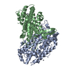
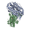
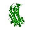
| Title | Ribonucleotide reductase Y730NO2Y modified R1 subunit of E. coli | |||||||||
 Components Components |
| |||||||||
 Keywords Keywords | OXIDOREDUCTASE / NUCLEOTIDE-BINDING / DNA REPLICATION / ALLOSTERIC ENZYME | |||||||||
| Function / homology |  Function and homology information Function and homology informationribonucleoside diphosphate metabolic process / 2'-deoxyribonucleotide biosynthetic process / nucleobase-containing small molecule interconversion / ribonucleoside-diphosphate reductase complex / ribonucleoside-diphosphate reductase / ribonucleoside-diphosphate reductase activity, thioredoxin disulfide as acceptor / deoxyribonucleotide biosynthetic process / protein folding chaperone / iron ion binding / ATP binding ...ribonucleoside diphosphate metabolic process / 2'-deoxyribonucleotide biosynthetic process / nucleobase-containing small molecule interconversion / ribonucleoside-diphosphate reductase complex / ribonucleoside-diphosphate reductase / ribonucleoside-diphosphate reductase activity, thioredoxin disulfide as acceptor / deoxyribonucleotide biosynthetic process / protein folding chaperone / iron ion binding / ATP binding / identical protein binding / cytoplasm / cytosol Similarity search - Function | |||||||||
| Biological species |  | |||||||||
| Method |  X-RAY DIFFRACTION / SYNCHROTRON / MOLECULAR REPLACEMENT / Resolution: 2.8 Å X-RAY DIFFRACTION / SYNCHROTRON / MOLECULAR REPLACEMENT / Resolution: 2.8 Å | |||||||||
 Authors Authors | Yokoyama, K. / Uhlin, U. / Stubbe, J. | |||||||||
 Citation Citation | Journal: J.Am.Chem.Soc. / Year: 2010 Title: Site-Specific Incorporation of 3-Nitrotyrosine as a Probe of Pk(A) Perturbation of Redox-Active Tyrosines in Ribonucleotide Reductase. Authors: Yokoyama, K. / Uhlin, U. / Stubbe, J. #1: Journal: Nature / Year: 1994Title: Structure of Ribonucleotide Reductase Protein R1. Authors: Uhlin, U. / Eklund, H. | |||||||||
| History |
|
- Structure visualization
| Structure viewer | Molecule: MolmilJmol/JSmol |
|---|
- Downloads & links
-Download
| PDBx/mmCIF format | 2xak.cif.gz | 445 KB | Display | PDBx/mmCIF format |
|---|---|---|---|---|
| PDB format | pdb2xak.ent.gz | 366.1 KB | Display | PDB format |
| PDBx/mmJSON format | 2xak.json.gz | Tree view | PDBx/mmJSON format | |
| Others |  Other downloads Other downloads |
-Validation report
| Arichive directory | https://data.pdbj.org/pub/pdb/validation_reports/xa/2xakftp://data.pdbj.org/pub/pdb/validation_reports/xa/2xak | HTTPS FTP |
|---|
-Related structure data
| Related structure data |  2x0xSC  2xapC  2xavC  2xawC  2xaxC  2xayC  2xazC S: Starting model for refinement C: citing same article ( |
|---|---|
| Similar structure data |


-Links
 PDBj
PDBj
- Assembly
| Deposited unit | 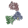
| ||||||||
|---|---|---|---|---|---|---|---|---|---|
| 1 | 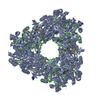
| ||||||||
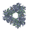
| 2 | x 6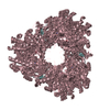
| ||||||||
| 3 | 
| ||||||||
| Unit cell |
| ||||||||
| Components on special symmetry positions |
|
-Components
| #1: Protein | Mass: 85922.086 Da / Num. of mol.: 3 / Fragment: RESIDUES 1-761 Source method: isolated from a genetically manipulated source Source: (gene. exp.) References: UniProt: P00452, ribonucleoside-diphosphate reductase #2: Protein/peptide | Mass: 2271.392 Da / Num. of mol.: 4 Fragment: RIBONUCLEOTIDE REDUCTASE R2-PEPTIDE, RESIDUES 357-376 Source method: obtained synthetically / Source: (synth.) References: UniProt: P69924, ribonucleoside-diphosphate reductase #3: Water | ChemComp-HOH / |  Mass: 18.015 Da / Num. of mol.: 439 / Source method: isolated from a natural source / Formula: H2O Mass: 18.015 Da / Num. of mol.: 439 / Source method: isolated from a natural source / Formula: H2ONonpolymer details | META-NITRO-TYROSINE (NIY): SITE SPECIFIC INCORPORAT | Sequence details | SITE SPECIFIC INCORPORAT | |
|---|
-Experimental details
-Experiment
| Experiment | Method: X-RAY DIFFRACTION / Number of used crystals: 1 |
|---|
- Sample preparation
| Crystal | Density Matthews: 3.05 Å3/Da / Density % sol: 54 % / Description: NONE |
|---|---|
| Crystal grow | pH: 6 Details: LITHIUM SULPHATE 1.5M, SODIUM CHLORIDE BUFFER PH 6. |
-Data collection
| Diffraction | Mean temperature: 100 K |
|---|---|
| Diffraction source | Source: SYNCHROTRON / Site: ESRF  / Beamline: ID14-2 / Wavelength: 0.933 / Beamline: ID14-2 / Wavelength: 0.933 |
| Detector | Type: ADSC CCD / Detector: CCD / Date: Jan 29, 2009 |
| Radiation | Protocol: SINGLE WAVELENGTH / Monochromatic (M) / Laue (L): M / Scattering type: x-ray |
| Radiation wavelength | Wavelength: 0.933 Å / Relative weight: 1 |
| Reflection | Resolution: 2.8→67.27 Å / Num. obs: 79929 / % possible obs: 100 % / Observed criterion σ(I): -3.7 / Redundancy: 9.71 % / Rmerge(I) obs: 0.14 / Net I/σ(I): 16.17 |
| Reflection shell | Resolution: 2.8→2.82 Å / Redundancy: 8.41 % / Rmerge(I) obs: 0.64 / Mean I/σ(I) obs: 3.35 / % possible all: 100 |
- Processing
| Software |
| ||||||||||||||||||||||||||||||||||||||||||||||||||||||||||||||||||||||||||||||||||||||||||||||||||||||||||||||||||||||||||||||||||||||||||||||||||||||||||||||||||||||||||||||||||||||
|---|---|---|---|---|---|---|---|---|---|---|---|---|---|---|---|---|---|---|---|---|---|---|---|---|---|---|---|---|---|---|---|---|---|---|---|---|---|---|---|---|---|---|---|---|---|---|---|---|---|---|---|---|---|---|---|---|---|---|---|---|---|---|---|---|---|---|---|---|---|---|---|---|---|---|---|---|---|---|---|---|---|---|---|---|---|---|---|---|---|---|---|---|---|---|---|---|---|---|---|---|---|---|---|---|---|---|---|---|---|---|---|---|---|---|---|---|---|---|---|---|---|---|---|---|---|---|---|---|---|---|---|---|---|---|---|---|---|---|---|---|---|---|---|---|---|---|---|---|---|---|---|---|---|---|---|---|---|---|---|---|---|---|---|---|---|---|---|---|---|---|---|---|---|---|---|---|---|---|---|---|---|---|---|
| Refinement | Method to determine structure: MOLECULAR REPLACEMENT Starting model: PDB ENTRY 2X0X Resolution: 2.8→169.031 Å / Cor.coef. Fo:Fc: 0.941 / Cor.coef. Fo:Fc free: 0.892 / SU B: 11.894 / SU ML: 0.234 / Cross valid method: THROUGHOUT / σ(F): 2 / ESU R: 1.296 / ESU R Free: 0.337 / Stereochemistry target values: MAXIMUM LIKELIHOOD Details: HYDROGENS HAVE BEEN ADDED IN THE RIDING POSITIONS. RESIDUES 268-273 ARE DISORDERED. THE 11-16 C-TERMINAL RESIDUES OF THE R2 PEPTIDE, CHAINS D, E, F BIND AT THE R2 BINDING-SITE OF THE R1 ...Details: HYDROGENS HAVE BEEN ADDED IN THE RIDING POSITIONS. RESIDUES 268-273 ARE DISORDERED. THE 11-16 C-TERMINAL RESIDUES OF THE R2 PEPTIDE, CHAINS D, E, F BIND AT THE R2 BINDING-SITE OF THE R1 MOLECULES, CHAINS A, B, C. THE THREE N-TERMINAL RESIDUES, UNIQUE CHAIN P, ARE SITUATED BETWEEN MOL A AND C. THIS LATTER BINDING HAS NO KNOWN BIOLOGICAL RELEVANCE BUT IS ESSENTIAL FOR CRYSTAL LATTICE FORMATION. WATERS CLOSE TO SER 625 MAY REPRESENT A SULPHATE ION.
| ||||||||||||||||||||||||||||||||||||||||||||||||||||||||||||||||||||||||||||||||||||||||||||||||||||||||||||||||||||||||||||||||||||||||||||||||||||||||||||||||||||||||||||||||||||||
| Solvent computation | Ion probe radii: 0.8 Å / VDW probe radii: 1.2 Å / Solvent model: MASK BULK SOLVENT | ||||||||||||||||||||||||||||||||||||||||||||||||||||||||||||||||||||||||||||||||||||||||||||||||||||||||||||||||||||||||||||||||||||||||||||||||||||||||||||||||||||||||||||||||||||||
| Displacement parameters | Biso mean: 36.914 Å2
| ||||||||||||||||||||||||||||||||||||||||||||||||||||||||||||||||||||||||||||||||||||||||||||||||||||||||||||||||||||||||||||||||||||||||||||||||||||||||||||||||||||||||||||||||||||||
| Refinement step | Cycle: LAST / Resolution: 2.8→169.031 Å
| ||||||||||||||||||||||||||||||||||||||||||||||||||||||||||||||||||||||||||||||||||||||||||||||||||||||||||||||||||||||||||||||||||||||||||||||||||||||||||||||||||||||||||||||||||||||
| Refine LS restraints |
|