 Movie
Movie Controller


+ Open data

- Basic information
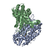
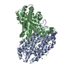
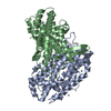
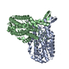
| Entry | Database: PDB / ID: 2av8 | ||||||
|---|---|---|---|---|---|---|---|
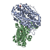
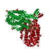
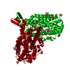
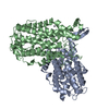
| Title | Y122F MUTANT OF RIBONUCLEOTIDE REDUCTASE FROM ESCHERICHIA COLI | ||||||
 Components Components | RIBONUCLEOTIDE REDUCTASE R2 | ||||||
 Keywords Keywords | OXIDOREDUCTASE / DNA REPLICATION | ||||||
| Function / homology |  Function and homology information Function and homology informationribonucleoside diphosphate metabolic process / 2'-deoxyribonucleotide biosynthetic process / nucleobase-containing small molecule interconversion / ribonucleoside-diphosphate reductase complex / ribonucleoside-diphosphate reductase / ribonucleoside-diphosphate reductase activity, thioredoxin disulfide as acceptor / deoxyribonucleotide biosynthetic process / iron ion binding / identical protein binding / cytoplasm / cytosol Similarity search - Function | ||||||
| Biological species |  | ||||||
| Method |  X-RAY DIFFRACTION / SYNCHROTRON / MOLECULAR REPLACEMENT / Resolution: 2.46 Å X-RAY DIFFRACTION / SYNCHROTRON / MOLECULAR REPLACEMENT / Resolution: 2.46 Å | ||||||
 Authors Authors | Han, S. / Arvai, A. / Tainer, J.A. | ||||||
 Citation Citation | Journal: Biochemistry / Year: 1998 Title: Characterization of Y122F R2 of Escherichia coli ribonucleotide reductase by time-resolved physical biochemical methods and X-ray crystallography. Authors: Tong, W. / Burdi, D. / Riggs-Gelasco, P. / Chen, S. / Edmondson, D. / Huynh, B.H. / Stubbe, J. / Han, S. / Arvai, A. / Tainer, J.A. #1: Journal: Science / Year: 1991Title: Mechanism of Assembly of the Tyrosyl Radical-Dinuclear Iron Cluster Cofactor of Ribonucleotide Reductase Authors: Bollinger Junior, J.M. / Edmondson, D.E. / Huynh, B.H. / Filley, J. / Norton, J.R. / Stubbe, J. | ||||||
| History |
|
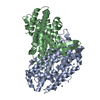
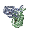
- Structure visualization
| Structure viewer | Molecule: MolmilJmol/JSmol |
|---|
- Downloads & links
-Download
| PDBx/mmCIF format | 2av8.cif.gz | 152.1 KB | Display | PDBx/mmCIF format |
|---|---|---|---|---|
| PDB format | pdb2av8.ent.gz | 120.4 KB | Display | PDB format |
| PDBx/mmJSON format | 2av8.json.gz | Tree view | PDBx/mmJSON format | |
| Others |  Other downloads Other downloads |
-Validation report
| Arichive directory | https://data.pdbj.org/pub/pdb/validation_reports/av/2av8ftp://data.pdbj.org/pub/pdb/validation_reports/av/2av8 | HTTPS FTP |
|---|
-Related structure data
| Related structure data |  1av8C  1ribS S: Starting model for refinement C: citing same article ( |
|---|---|
| Similar structure data |


-Links
 PDBj
PDBj


- Assembly
| Deposited unit | 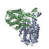
| ||||||||
|---|---|---|---|---|---|---|---|---|---|
| 1 |
| ||||||||
| Unit cell |
| ||||||||
| Noncrystallographic symmetry (NCS) | NCS oper: (Code: given Matrix: (0.999097, 0.037832, 0.019338), Vector: |
-Components
| #1: Protein | Mass: 39587.883 Da / Num. of mol.: 2 / Mutation: Y122F Source method: isolated from a genetically manipulated source Source: (gene. exp.) References: UniProt: P69924, ribonucleoside-diphosphate reductase #2: Chemical | ChemComp-FE2 / |   Mass: 55.845 Da / Num. of mol.: 1 / Source method: obtained synthetically / Formula: Fe Mass: 55.845 Da / Num. of mol.: 1 / Source method: obtained synthetically / Formula: Fe#3: Chemical |   Mass: 127.689 Da / Num. of mol.: 2 / Source method: obtained synthetically / Formula: Fe2O Mass: 127.689 Da / Num. of mol.: 2 / Source method: obtained synthetically / Formula: Fe2O#4: Water | ChemComp-HOH / |  Mass: 18.015 Da / Num. of mol.: 204 / Source method: isolated from a natural source / Formula: H2O Mass: 18.015 Da / Num. of mol.: 204 / Source method: isolated from a natural source / Formula: H2O |
|---|
-Experimental details
-Experiment
| Experiment | Method: X-RAY DIFFRACTION / Number of used crystals: 1 |
|---|
- Sample preparation
| Crystal | Density Matthews: 3.5 Å3/Da / Density % sol: 65 % | ||||||||||||||||||||||||||||||
|---|---|---|---|---|---|---|---|---|---|---|---|---|---|---|---|---|---|---|---|---|---|---|---|---|---|---|---|---|---|---|---|
| Crystal grow | pH: 7.6 Details: PROTEIN WAS CRYSTALLIZED FROM 80% SATURATED NACL, NEAR PHYSIOLOGICAL PH (PH 7.6) | ||||||||||||||||||||||||||||||
| Crystal grow | *PLUS Method: vapor diffusion | ||||||||||||||||||||||||||||||
| Components of the solutions | *PLUS
|
-Data collection
| Diffraction | Mean temperature: 103 K |
|---|---|
| Diffraction source | Source: SYNCHROTRON / Site: SSRL  / Beamline: BL7-1 / Wavelength: 1.08 / Beamline: BL7-1 / Wavelength: 1.08 |
| Detector | Detector: IMAGE PLATE / Date: Aug 1, 1996 |
| Radiation | Monochromatic (M) / Laue (L): M / Scattering type: x-ray |
| Radiation wavelength | Wavelength: 1.08 Å / Relative weight: 1 |
| Reflection | Resolution: 2.46→100 Å / Num. obs: 39923 / % possible obs: 95 % / Observed criterion σ(I): -3 / Rsym value: 0.058 / Net I/σ(I): 14.4 |
| Reflection shell | Resolution: 2.46→2.55 Å / Redundancy: 2.55 % / Mean I/σ(I) obs: 2.4 / Rsym value: 0.302 / % possible all: 82.7 |
| Reflection | *PLUS Rmerge(I) obs: 0.056 |
| Reflection shell | *PLUS % possible obs: 83 % |
- Processing
| Software |
| ||||||||||||||||||||||||||||||||||||||||||||||||||||||||||||
|---|---|---|---|---|---|---|---|---|---|---|---|---|---|---|---|---|---|---|---|---|---|---|---|---|---|---|---|---|---|---|---|---|---|---|---|---|---|---|---|---|---|---|---|---|---|---|---|---|---|---|---|---|---|---|---|---|---|---|---|---|---|
| Refinement | Method to determine structure: MOLECULAR REPLACEMENT Starting model: 1RIB Resolution: 2.46→7 Å / Cross valid method: THROUGHOUT / σ(F): 1
| ||||||||||||||||||||||||||||||||||||||||||||||||||||||||||||
| Refinement step | Cycle: LAST / Resolution: 2.46→7 Å
| ||||||||||||||||||||||||||||||||||||||||||||||||||||||||||||
| Refine LS restraints |
| ||||||||||||||||||||||||||||||||||||||||||||||||||||||||||||
| Software | *PLUS Name: X-PLOR / Version: 3.8 / Classification: refinement | ||||||||||||||||||||||||||||||||||||||||||||||||||||||||||||
| Refine LS restraints | *PLUS
|
