 Movie
Movie Controller

[English] 日本語
 Yorodumi
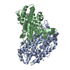
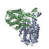
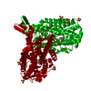
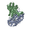
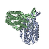
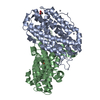
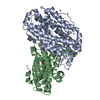
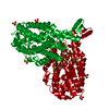
- PDB-1pj0: RIBONUCLEOTIDE REDUCTASE R2-D84E/W48F MUTANT SOAKED WITH FERROUS ... -
+ Open data

- Basic information
| Entry | Database: PDB / ID: 1pj0 | ||||||
|---|---|---|---|---|---|---|---|
| Title | RIBONUCLEOTIDE REDUCTASE R2-D84E/W48F MUTANT SOAKED WITH FERROUS IONS AT NEUTRAL PH | ||||||
 Components Components | Ribonucleoside-diphosphate reductase 1 beta chain | ||||||
 Keywords Keywords | OXIDOREDUCTASE / four helix bundle / diferrous cluster | ||||||
| Function / homology |  Function and homology information Function and homology informationribonucleoside diphosphate metabolic process / 2'-deoxyribonucleotide biosynthetic process / nucleobase-containing small molecule interconversion / ribonucleoside-diphosphate reductase complex / ribonucleoside-diphosphate reductase / ribonucleoside-diphosphate reductase activity, thioredoxin disulfide as acceptor / deoxyribonucleotide biosynthetic process / iron ion binding / identical protein binding / cytosol / cytoplasm Similarity search - Function | ||||||
| Biological species |  | ||||||
| Method |  X-RAY DIFFRACTION / SYNCHROTRON / MOLECULAR REPLACEMENT / Resolution: 1.9 Å X-RAY DIFFRACTION / SYNCHROTRON / MOLECULAR REPLACEMENT / Resolution: 1.9 Å | ||||||
 Authors Authors | Voegtli, W.C. / Sommerhalter, M. / Saleh, L. / Baldwin, J. / Bollinger Jr., J.M. / Rosenzweig, A.C. | ||||||
 Citation Citation | Journal: J.Am.Chem.Soc. / Year: 2003 Title: Variable coordination geometries at the diiron(II) active site of ribonucleotide reductase R2. Authors: Voegtli, W.C. / Sommerhalter, M. / Saleh, L. / Baldwin, J. / Bollinger Jr., J.M. / Rosenzweig, A.C. | ||||||
| History |
| ||||||
| Remark 999 | SEQUENCE THE CONFLICTS ARE DUE TO A MODELLING ERROR IN THE INITIAL MODEL. |
- Structure visualization
| Structure viewer | Molecule: MolmilJmol/JSmol |
|---|
- Downloads & links
-Download
| PDBx/mmCIF format | 1pj0.cif.gz | 154.9 KB | Display | PDBx/mmCIF format |
|---|---|---|---|---|
| PDB format | pdb1pj0.ent.gz | 122.3 KB | Display | PDB format |
| PDBx/mmJSON format | 1pj0.json.gz | Tree view | PDBx/mmJSON format | |
| Others |  Other downloads Other downloads |
-Validation report
| Arichive directory | https://data.pdbj.org/pub/pdb/validation_reports/pj/1pj0ftp://data.pdbj.org/pub/pdb/validation_reports/pj/1pj0 | HTTPS FTP |
|---|
-Related structure data
| Related structure data |  1piyC  1pizC  1pj1C  1pm2C  1r65C  1pimS S: Starting model for refinement C: citing same article ( |
|---|---|
| Similar structure data |


-Links
 PDBj
PDBj


- Assembly
| Deposited unit | 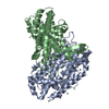
| ||||||||
|---|---|---|---|---|---|---|---|---|---|
| 1 |
| ||||||||
| Unit cell |
| ||||||||
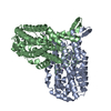
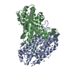
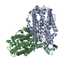
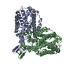
| Details | Tetramer of two alpha and two beta subunits |
-Components
| #1: Protein | Mass: 43387.824 Da / Num. of mol.: 2 / Mutation: D84E, W48F Source method: isolated from a genetically manipulated source Source: (gene. exp.) References: UniProt: P69924, ribonucleoside-diphosphate reductase #2: Chemical | ChemComp-FE /   Mass: 55.845 Da / Num. of mol.: 4 / Source method: obtained synthetically / Formula: Fe Mass: 55.845 Da / Num. of mol.: 4 / Source method: obtained synthetically / Formula: Fe#3: Chemical | ChemComp-HG /   Mass: 200.590 Da / Num. of mol.: 9 / Source method: obtained synthetically / Formula: Hg Mass: 200.590 Da / Num. of mol.: 9 / Source method: obtained synthetically / Formula: Hg#4: Water | ChemComp-HOH / |  Mass: 18.015 Da / Num. of mol.: 254 / Source method: isolated from a natural source / Formula: H2O Mass: 18.015 Da / Num. of mol.: 254 / Source method: isolated from a natural source / Formula: H2OHas protein modification | Y | |
|---|
-Experimental details
-Experiment
| Experiment | Method: X-RAY DIFFRACTION / Number of used crystals: 1 |
|---|
- Sample preparation
| Crystal | Density Matthews: 2.06 Å3/Da / Density % sol: 40.33 % | |||||||||||||||||||||||||||||||||||||||||||||||||||||||||||||||
|---|---|---|---|---|---|---|---|---|---|---|---|---|---|---|---|---|---|---|---|---|---|---|---|---|---|---|---|---|---|---|---|---|---|---|---|---|---|---|---|---|---|---|---|---|---|---|---|---|---|---|---|---|---|---|---|---|---|---|---|---|---|---|---|---|
| Crystal grow | Temperature: 310 K / Method: vapor diffusion, hanging drop / pH: 6 Details: 20% PEG 4000, 200mM NaCl, 50mM MES, 0.3% dioxane, pH 6.0, VAPOR DIFFUSION, HANGING DROP, temperature 310K | |||||||||||||||||||||||||||||||||||||||||||||||||||||||||||||||
| Crystal grow | *PLUS Temperature: 37 ℃ / pH: 7.6 / Method: vapor diffusion, hanging drop | |||||||||||||||||||||||||||||||||||||||||||||||||||||||||||||||
| Components of the solutions | *PLUS
|
-Data collection
| Diffraction | Mean temperature: 100 K |
|---|---|
| Diffraction source | Source: SYNCHROTRON / Site: SSRL  / Beamline: BL9-1 / Wavelength: 0.97 Å / Beamline: BL9-1 / Wavelength: 0.97 Å |
| Detector | Type: MARRESEARCH / Detector: IMAGE PLATE / Date: Jun 30, 2001 |
| Radiation | Protocol: SINGLE WAVELENGTH / Monochromatic (M) / Laue (L): M / Scattering type: x-ray |
| Radiation wavelength | Wavelength: 0.97 Å / Relative weight: 1 |
| Reflection | Resolution: 1.9→20 Å / Num. all: 57203 / Num. obs: 54370 / % possible obs: 95 % / Observed criterion σ(F): 1 / Observed criterion σ(I): 1 / Biso Wilson estimate: 15.4 Å2 |
| Reflection shell | Resolution: 1.9→2.07 Å / % possible all: 89 |
| Reflection | *PLUS Lowest resolution: 25 Å / Num. obs: 55199 / % possible obs: 98.1 % / Num. measured all: 556805 / Rmerge(I) obs: 0.066 |
| Reflection shell | *PLUS % possible obs: 93.8 % / Rmerge(I) obs: 0.324 / Mean I/σ(I) obs: 2.5 |
- Processing
| Software |
| ||||||||||||||||||||||||||||||||||||
|---|---|---|---|---|---|---|---|---|---|---|---|---|---|---|---|---|---|---|---|---|---|---|---|---|---|---|---|---|---|---|---|---|---|---|---|---|---|
| Refinement | Method to determine structure: MOLECULAR REPLACEMENT Starting model: PDB ENTRY 1PIM Resolution: 1.9→19.32 Å / Rfactor Rfree error: 0.005 / Data cutoff high absF: 592822.39 / Data cutoff high rms absF: 592822.39 / Data cutoff low absF: 0 / Isotropic thermal model: RESTRAINED / Cross valid method: THROUGHOUT / σ(F): 0 / Stereochemistry target values: Engh & Huber
| ||||||||||||||||||||||||||||||||||||
| Solvent computation | Solvent model: FLAT MODEL / Bsol: 47.7607 Å2 / ksol: 0.391806 e/Å3 | ||||||||||||||||||||||||||||||||||||
| Displacement parameters | Biso mean: 23.4 Å2
| ||||||||||||||||||||||||||||||||||||
| Refine analyze |
| ||||||||||||||||||||||||||||||||||||
| Refinement step | Cycle: LAST / Resolution: 1.9→19.32 Å
| ||||||||||||||||||||||||||||||||||||
| Refine LS restraints |
| ||||||||||||||||||||||||||||||||||||
| LS refinement shell | Resolution: 1.9→2.02 Å / Rfactor Rfree error: 0.013 / Total num. of bins used: 6
| ||||||||||||||||||||||||||||||||||||
| Xplor file |
| ||||||||||||||||||||||||||||||||||||
| Refinement | *PLUS Lowest resolution: 25 Å / % reflection Rfree: 4.8 % / Rfactor Rwork: 0.205 | ||||||||||||||||||||||||||||||||||||
| Solvent computation | *PLUS | ||||||||||||||||||||||||||||||||||||
| Displacement parameters | *PLUS | ||||||||||||||||||||||||||||||||||||
| Refine LS restraints | *PLUS
|
