 Movie
Movie Controller

[English] 日本語
 Yorodumi
- PDB-1jqc: Mn substituted Ribonucleotide reductase R2 from E. Coli oxidized ... -
+ Open data

- Basic information
| Entry | Database: PDB / ID: 1jqc | ||||||
|---|---|---|---|---|---|---|---|
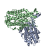
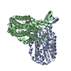
| Title | Mn substituted Ribonucleotide reductase R2 from E. Coli oxidized by hydrogen peroxide and hydroxylamine | ||||||
 Components Components | Protein R2 of ribonucleotide reductase | ||||||
 Keywords Keywords | OXIDOREDUCTASE / Ribonucleotide reductase R2 / Radical protein / Mn substituted / oxidized By H2O2/NH2OH | ||||||
| Function / homology |  Function and homology information Function and homology informationribonucleoside diphosphate metabolic process / 2'-deoxyribonucleotide biosynthetic process / nucleobase-containing small molecule interconversion / ribonucleoside-diphosphate reductase complex / ribonucleoside-diphosphate reductase / ribonucleoside-diphosphate reductase activity, thioredoxin disulfide as acceptor / deoxyribonucleotide biosynthetic process / iron ion binding / identical protein binding / cytoplasm / cytosol Similarity search - Function | ||||||
| Biological species |  | ||||||
| Method |  X-RAY DIFFRACTION / SYNCHROTRON / MOLECULAR REPLACEMENT / Resolution: 1.61 Å X-RAY DIFFRACTION / SYNCHROTRON / MOLECULAR REPLACEMENT / Resolution: 1.61 Å | ||||||
 Authors Authors | Hogbom, M. / Andersson, M.E. / Nordlund, P. | ||||||
 Citation Citation | Journal: J.Biol.Inorg.Chem. / Year: 2001 Title: Crystal structures of oxidized dinuclear manganese centres in Mn-substituted class I ribonucleotide reductase from Escherichia coli: carboxylate shifts with implications for O2 activation and radical generation. Authors: Hogbom, M. / Andersson, M.E. / Nordlund, P. | ||||||
| History |
|
- Structure visualization
| Structure viewer | Molecule: MolmilJmol/JSmol |
|---|
- Downloads & links
-Download
| PDBx/mmCIF format | 1jqc.cif.gz | 169.7 KB | Display | PDBx/mmCIF format |
|---|---|---|---|---|
| PDB format | pdb1jqc.ent.gz | 133.1 KB | Display | PDB format |
| PDBx/mmJSON format | 1jqc.json.gz | Tree view | PDBx/mmJSON format | |
| Others |  Other downloads Other downloads |
-Validation report
| Arichive directory | https://data.pdbj.org/pub/pdb/validation_reports/jq/1jqcftp://data.pdbj.org/pub/pdb/validation_reports/jq/1jqc | HTTPS FTP |
|---|
-Related structure data


-Links
 PDBj
PDBj


- Assembly
| Deposited unit | 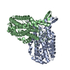
| ||||||||
|---|---|---|---|---|---|---|---|---|---|
| 1 |
| ||||||||
| Unit cell |
|
-Components
| #1: Protein | Mass: 43426.863 Da / Num. of mol.: 2 Source method: isolated from a genetically manipulated source Source: (gene. exp.) References: UniProt: P69924, ribonucleoside-diphosphate reductase #2: Chemical | ChemComp-MN /   Mass: 54.938 Da / Num. of mol.: 4 / Source method: obtained synthetically / Formula: Mn Mass: 54.938 Da / Num. of mol.: 4 / Source method: obtained synthetically / Formula: Mn#3: Chemical | ChemComp-HG /   Mass: 200.590 Da / Num. of mol.: 13 / Source method: obtained synthetically / Formula: Hg Mass: 200.590 Da / Num. of mol.: 13 / Source method: obtained synthetically / Formula: Hg#4: Water | ChemComp-HOH / |  Mass: 18.015 Da / Num. of mol.: 726 / Source method: isolated from a natural source / Formula: H2O Mass: 18.015 Da / Num. of mol.: 726 / Source method: isolated from a natural source / Formula: H2O |
|---|
-Experimental details
-Experiment
| Experiment | Method: X-RAY DIFFRACTION / Number of used crystals: 1 |
|---|
- Sample preparation
| Crystal | Density Matthews: 2.05 Å3/Da / Density % sol: 40.1 % | |||||||||||||||||||||||||||||||||||||||||||||||||
|---|---|---|---|---|---|---|---|---|---|---|---|---|---|---|---|---|---|---|---|---|---|---|---|---|---|---|---|---|---|---|---|---|---|---|---|---|---|---|---|---|---|---|---|---|---|---|---|---|---|---|
| Crystal grow | Temperature: 298 K / Method: vapor diffusion, hanging drop / pH: 6 Details: peg 4000, MES, EMTS, sodium chloride, pH 6.0, VAPOR DIFFUSION, HANGING DROP, temperature 298K | |||||||||||||||||||||||||||||||||||||||||||||||||
| Crystal grow | *PLUS Temperature: 20 ℃ / Details: Nordlund, P., (1989) FEBS Lett., 258, 251. | |||||||||||||||||||||||||||||||||||||||||||||||||
| Components of the solutions | *PLUS
|
-Data collection
| Diffraction | Mean temperature: 100 K |
|---|---|
| Diffraction source | Source: SYNCHROTRON / Site: ESRF  / Beamline: BM1A / Wavelength: 0.88 Å / Beamline: BM1A / Wavelength: 0.88 Å |
| Detector | Type: MARRESEARCH / Detector: IMAGE PLATE / Date: Feb 7, 1999 |
| Radiation | Monochromator: SAGITALLY FOCUSED Si(111) / Protocol: SINGLE WAVELENGTH / Monochromatic (M) / Laue (L): M / Scattering type: x-ray |
| Radiation wavelength | Wavelength: 0.88 Å / Relative weight: 1 |
| Reflection | Resolution: 1.61→29 Å / Num. all: 91243 / Num. obs: 91243 / % possible obs: 97.8 % / Observed criterion σ(F): 0 / Observed criterion σ(I): 0 / Redundancy: 3.9 % / Rmerge(I) obs: 0.065 |
| Reflection shell | Resolution: 1.61→1.71 Å / % possible all: 95.3 |
| Reflection | *PLUS Lowest resolution: 29 Å |
| Reflection shell | *PLUS % possible obs: 99.3 % / Redundancy: 3.9 % / Rmerge(I) obs: 0.286 |
- Processing
| Software |
| |||||||||||||||||||||||||
|---|---|---|---|---|---|---|---|---|---|---|---|---|---|---|---|---|---|---|---|---|---|---|---|---|---|---|
| Refinement | Method to determine structure: MOLECULAR REPLACEMENT / Resolution: 1.61→29 Å / σ(F): 0 / Stereochemistry target values: Engh & Huber
| |||||||||||||||||||||||||
| Refinement step | Cycle: LAST / Resolution: 1.61→29 Å
| |||||||||||||||||||||||||
| Refine LS restraints |
| |||||||||||||||||||||||||
| Refinement | *PLUS Lowest resolution: 29 Å / σ(F): 0 / % reflection Rfree: 5 % / Rfactor obs: 0.176 | |||||||||||||||||||||||||
| Solvent computation | *PLUS | |||||||||||||||||||||||||
| Displacement parameters | *PLUS | |||||||||||||||||||||||||
| Refine LS restraints | *PLUS Type: c_angle_deg / Dev ideal: 1.8 |
