 Movie
Movie Controller

[English] 日本語
 Yorodumi
- PDB-5r1r: RIBONUCLEOTIDE REDUCTASE E441A MUTANT R1 PROTEIN FROM ESCHERICHIA COLI -
+ Open data

- Basic information
| Entry | Database: PDB / ID: 5r1r | ||||||
|---|---|---|---|---|---|---|---|
| Title | RIBONUCLEOTIDE REDUCTASE E441A MUTANT R1 PROTEIN FROM ESCHERICHIA COLI | ||||||
 Components Components |
| ||||||
 Keywords Keywords | COMPLEX (OXIDOREDUCTASE/PEPTIDE) / RIBONUCLEOTIDE REDUCTASE / DEOXYRIBONUCLEOTIDE SYNTHESIS / RADICAL CHEMISTRY / ALLOSTERIC REGULATION / SPECIFICITY / COMPLEX (OXIDOREDUCTASE-PEPTIDE) / COMPLEX (OXIDOREDUCTASE-PEPTIDE) complex | ||||||
| Function / homology |  Function and homology information Function and homology informationribonucleoside diphosphate metabolic process / 2'-deoxyribonucleotide biosynthetic process / nucleobase-containing small molecule interconversion / ribonucleoside-diphosphate reductase complex / ribonucleoside-diphosphate reductase / ribonucleoside-diphosphate reductase activity, thioredoxin disulfide as acceptor / deoxyribonucleotide biosynthetic process / protein folding chaperone / iron ion binding / ATP binding ...ribonucleoside diphosphate metabolic process / 2'-deoxyribonucleotide biosynthetic process / nucleobase-containing small molecule interconversion / ribonucleoside-diphosphate reductase complex / ribonucleoside-diphosphate reductase / ribonucleoside-diphosphate reductase activity, thioredoxin disulfide as acceptor / deoxyribonucleotide biosynthetic process / protein folding chaperone / iron ion binding / ATP binding / identical protein binding / cytoplasm / cytosol Similarity search - Function | ||||||
| Biological species |  | ||||||
| Method |  X-RAY DIFFRACTION / SYNCHROTRON / RIGID BODY / Resolution: 3.1 Å X-RAY DIFFRACTION / SYNCHROTRON / RIGID BODY / Resolution: 3.1 Å | ||||||
 Authors Authors | Eriksson, M. / Eklund, H. | ||||||
 Citation Citation | Journal: J.Biol.Chem. / Year: 1997 Title: A new mechanism-based radical intermediate in a mutant R1 protein affecting the catalytically essential Glu441 in Escherichia coli ribonucleotide reductase. Authors: Persson, A.L. / Eriksson, M. / Katterle, B. / Potsch, S. / Sahlin, M. / Sjoberg, B.M. #1: Journal: Structure / Year: 1997Title: Binding of Allosteric Effectors to Ribonucleotide Reductase Protein R1: Reduction of Active-Site Cysteines Promotes Substrate Binding Authors: Eriksson, M. / Uhlin, U. / Ramaswamy, S. / Ekberg, M. / Regnstrom, K. / Sjoberg, B.M. / Eklund, H. #2: Journal: Nature / Year: 1994Title: Structure of Ribonucleotide Reductase Protein R1 Authors: Uhlin, U. / Eklund, H. | ||||||
| History |
|
- Structure visualization
| Structure viewer | Molecule: MolmilJmol/JSmol |
|---|
- Downloads & links
-Download
| PDBx/mmCIF format | 5r1r.cif.gz | 441.5 KB | Display | PDBx/mmCIF format |
|---|---|---|---|---|
| PDB format | pdb5r1r.ent.gz | 364.3 KB | Display | PDB format |
| PDBx/mmJSON format | 5r1r.json.gz | Tree view | PDBx/mmJSON format | |
| Others |  Other downloads Other downloads |
-Validation report
| Arichive directory | https://data.pdbj.org/pub/pdb/validation_reports/r1/5r1rftp://data.pdbj.org/pub/pdb/validation_reports/r1/5r1r | HTTPS FTP |
|---|
-Related structure data
| Related structure data |  6r1rC  7r1rC  1rlrS S: Starting model for refinement C: citing same article ( |
|---|---|
| Similar structure data |


-Links
 PDBj
PDBj
- Assembly
| Deposited unit | 
| ||||||||||||
|---|---|---|---|---|---|---|---|---|---|---|---|---|---|
| 1 | 
| ||||||||||||
| 2 | 
| ||||||||||||
| 3 | 
| ||||||||||||
| 4 | 
| ||||||||||||
| 5 | x 6
| ||||||||||||
| 6 | 
| ||||||||||||
| 7 | 
| ||||||||||||
| 8 | 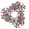
| ||||||||||||
| 9 | 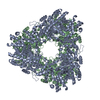
| ||||||||||||
| 10 | 
| ||||||||||||
| 11 | 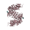
| ||||||||||||
| 12 | 
| ||||||||||||
| Unit cell |
| ||||||||||||
| Noncrystallographic symmetry (NCS) | NCS oper:
|
-Components
| #1: Protein | Mass: 85819.055 Da / Num. of mol.: 3 / Mutation: E441A Source method: isolated from a genetically manipulated source Source: (gene. exp.) References: UniProt: P00452, ribonucleoside-diphosphate reductase #2: Protein/peptide | Mass: 2271.392 Da / Num. of mol.: 4 / Fragment: C-TERMINAL PORTION, 20 RESIDUES Source method: isolated from a genetically manipulated source Source: (gene. exp.) #3: Water | ChemComp-HOH / |  Mass: 18.015 Da / Num. of mol.: 90 / Source method: isolated from a natural source / Formula: H2O Mass: 18.015 Da / Num. of mol.: 90 / Source method: isolated from a natural source / Formula: H2OHas protein modification | Y | |
|---|
-Experimental details
-Experiment
| Experiment | Method: X-RAY DIFFRACTION / Number of used crystals: 1 |
|---|
- Sample preparation
| Crystal | Density Matthews: 3.02 Å3/Da / Density % sol: 56 % | |||||||||||||||||||||||||
|---|---|---|---|---|---|---|---|---|---|---|---|---|---|---|---|---|---|---|---|---|---|---|---|---|---|---|
| Crystal grow | pH: 6 Details: PROTEIN WAS CRYSTALLIZED FROM 1.7 M LITHIUM SULFATE, AND 10 MM MAGNESIUM SULFATE IN 25 MM CITRATE BUFFER AT PH 6.0 THE PROTEIN SOLUTION CONTAINED 17 MG/ML R1 PROTEIN, 20 FOLD EXCESS OF A 20- ...Details: PROTEIN WAS CRYSTALLIZED FROM 1.7 M LITHIUM SULFATE, AND 10 MM MAGNESIUM SULFATE IN 25 MM CITRATE BUFFER AT PH 6.0 THE PROTEIN SOLUTION CONTAINED 17 MG/ML R1 PROTEIN, 20 FOLD EXCESS OF A 20-RESIDUE PEPTIDE CORRESPONDING TO THE C-TERMINUS OF THE R2 SUBUNIT AND IS ESSENTIAL FOR CRYSTALLIZATION | |||||||||||||||||||||||||
| Crystal grow | *PLUS Method: vapor diffusion, hanging drop | |||||||||||||||||||||||||
| Components of the solutions | *PLUS
|
-Data collection
| Diffraction | Mean temperature: 100 K |
|---|---|
| Diffraction source | Source: SYNCHROTRON / Site: NSLS  / Type: NSLS / Wavelength: 1.5418 / Type: NSLS / Wavelength: 1.5418 |
| Detector | Type: RIGAKU RAXIS IIC / Detector: IMAGE PLATE / Date: Nov 1, 1995 |
| Radiation | Monochromatic (M) / Laue (L): M / Scattering type: x-ray |
| Radiation wavelength | Wavelength: 1.5418 Å / Relative weight: 1 |
| Reflection | Resolution: 3.1→40 Å / Num. obs: 53031 / % possible obs: 90.9 % / Observed criterion σ(I): -3 / Redundancy: 2.8 % / Rmerge(I) obs: 0.071 / Rsym value: 0.071 / Net I/σ(I): 13.2 |
| Reflection shell | Resolution: 3.1→3.2 Å / Redundancy: 2.4 % / Rmerge(I) obs: 0.29 / Mean I/σ(I) obs: 2.8 / Rsym value: 0.29 / % possible all: 89.9 |
- Processing
| Software |
| ||||||||||||||||||||
|---|---|---|---|---|---|---|---|---|---|---|---|---|---|---|---|---|---|---|---|---|---|
| Refinement | Method to determine structure: RIGID BODY Starting model: PDB ENTRY 1RLR Resolution: 3.1→20 Å / Cross valid method: THROUGHOUT / σ(F): 0
| ||||||||||||||||||||
| Refinement step | Cycle: LAST / Resolution: 3.1→20 Å
| ||||||||||||||||||||
| Software | *PLUS Name: REFMAC / Classification: refinement | ||||||||||||||||||||
| Refinement | *PLUS Num. reflection all: 52838 | ||||||||||||||||||||
| Solvent computation | *PLUS | ||||||||||||||||||||
| Displacement parameters | *PLUS | ||||||||||||||||||||
| Refine LS restraints | *PLUS
|
