 Movie
Movie Controller

[English] 日本語
 Yorodumi
- PDB-4r1r: RIBONUCLEOTIDE REDUCTASE R1 PROTEIN WITH SUBSTRATE, GDP AND EFFEC... -
+ Open data

- Basic information
| Entry | Database: PDB / ID: 4r1r | ||||||
|---|---|---|---|---|---|---|---|
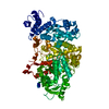
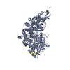
| Title | RIBONUCLEOTIDE REDUCTASE R1 PROTEIN WITH SUBSTRATE, GDP AND EFFECTOR DTTP FROM ESCHERICHIA COLI | ||||||
 Components Components |
| ||||||
 Keywords Keywords | COMPLEX (OXIDOREDUCTASE/PEPTIDE) / RIBONUCLEOTIDE REDUCTASE / DEOXYRIBONUCLEOTIDE SYNTHESIS / RADICAL CHEMISTRY / ALLOSTERIC REGULATION / SPECIFICITY / COMPLEX (OXIDOREDUCTASE-PEPTIDE) / COMPLEX (OXIDOREDUCTASE-PEPTIDE) complex | ||||||
| Function / homology |  Function and homology information Function and homology informationribonucleoside diphosphate metabolic process / 2'-deoxyribonucleotide biosynthetic process / nucleobase-containing small molecule interconversion / ribonucleoside-diphosphate reductase complex / ribonucleoside-diphosphate reductase / ribonucleoside-diphosphate reductase activity, thioredoxin disulfide as acceptor / deoxyribonucleotide biosynthetic process / protein folding chaperone / iron ion binding / ATP binding ...ribonucleoside diphosphate metabolic process / 2'-deoxyribonucleotide biosynthetic process / nucleobase-containing small molecule interconversion / ribonucleoside-diphosphate reductase complex / ribonucleoside-diphosphate reductase / ribonucleoside-diphosphate reductase activity, thioredoxin disulfide as acceptor / deoxyribonucleotide biosynthetic process / protein folding chaperone / iron ion binding / ATP binding / identical protein binding / cytosol / cytoplasm Similarity search - Function | ||||||
| Biological species |  | ||||||
| Method |  X-RAY DIFFRACTION / RIGID BODY / Resolution: 3.2 Å X-RAY DIFFRACTION / RIGID BODY / Resolution: 3.2 Å | ||||||
 Authors Authors | Eriksson, M. / Eklund, H. | ||||||
 Citation Citation | Journal: Structure / Year: 1997 Title: Binding of allosteric effectors to ribonucleotide reductase protein R1: reduction of active-site cysteines promotes substrate binding. Authors: Eriksson, M. / Uhlin, U. / Ramaswamy, S. / Ekberg, M. / Regnstrom, K. / Sjoberg, B.M. / Eklund, H. #1: Journal: Nature / Year: 1994Title: Structure of Ribonucleotide Reductase Protein R1 Authors: Uhlin, U. / Eklund, H. | ||||||
| History |
|
- Structure visualization
| Structure viewer | Molecule: MolmilJmol/JSmol |
|---|
- Downloads & links
-Download
| PDBx/mmCIF format | 4r1r.cif.gz | 443.2 KB | Display | PDBx/mmCIF format |
|---|---|---|---|---|
| PDB format | pdb4r1r.ent.gz | 363.6 KB | Display | PDB format |
| PDBx/mmJSON format | 4r1r.json.gz | Tree view | PDBx/mmJSON format | |
| Others |  Other downloads Other downloads |
-Validation report
| Arichive directory | https://data.pdbj.org/pub/pdb/validation_reports/r1/4r1rftp://data.pdbj.org/pub/pdb/validation_reports/r1/4r1r | HTTPS FTP |
|---|
-Related structure data


-Links
 PDBj
PDBj
- Assembly
| Deposited unit | 
| ||||||||||||
|---|---|---|---|---|---|---|---|---|---|---|---|---|---|
| 1 | 
| ||||||||||||
| 2 | 
| ||||||||||||
| 3 | 
| ||||||||||||
| 4 | 
| ||||||||||||
| 5 | x 6
| ||||||||||||
| 6 | 
| ||||||||||||
| 7 | 
| ||||||||||||
| 8 | 
| ||||||||||||
| 9 | 
| ||||||||||||
| 10 | 
| ||||||||||||
| Unit cell |
| ||||||||||||
| Noncrystallographic symmetry (NCS) | NCS oper:
|
-Components
| #1: Protein | Mass: 85845.023 Da / Num. of mol.: 3 / Mutation: C292A Source method: isolated from a genetically manipulated source Source: (gene. exp.) References: UniProt: P00452, ribonucleoside-diphosphate reductase #2: Protein/peptide | Mass: 2271.392 Da / Num. of mol.: 4 / Fragment: C-TERMINAL PORTION, 20 RESIDUES Source method: isolated from a genetically manipulated source Source: (gene. exp.) #3: Chemical |   Mass: 482.168 Da / Num. of mol.: 3 / Source method: obtained synthetically / Formula: C10H17N2O14P3 Mass: 482.168 Da / Num. of mol.: 3 / Source method: obtained synthetically / Formula: C10H17N2O14P3#4: Chemical |   Type: RNA linking / Mass: 443.201 Da / Num. of mol.: 3 / Source method: obtained synthetically / Formula: C10H15N5O11P2 / Comment: GDP, energy-carrying molecule*YM Type: RNA linking / Mass: 443.201 Da / Num. of mol.: 3 / Source method: obtained synthetically / Formula: C10H15N5O11P2 / Comment: GDP, energy-carrying molecule*YM |
|---|
-Experimental details
-Experiment
| Experiment | Method: X-RAY DIFFRACTION / Number of used crystals: 1 |
|---|
- Sample preparation
| Crystal | Density Matthews: 3.04 Å3/Da / Density % sol: 56.5 % | ||||||||||||||||||||||||||||||||||||
|---|---|---|---|---|---|---|---|---|---|---|---|---|---|---|---|---|---|---|---|---|---|---|---|---|---|---|---|---|---|---|---|---|---|---|---|---|---|
| Crystal grow | pH: 6 Details: PROTEIN WAS CRYSTALLIZED FROM 1.7 M LITHIUM SULFATE, AND 10 MM MAGNESIUM SULFATE IN 25 MM CITRATE BUFFER AT PH 6.0. THE PROTEIN SOLUTION CONTAINED 17 MG/ML R1 PROTEIN, 20-FOLD EXCESS FO A 20- ...Details: PROTEIN WAS CRYSTALLIZED FROM 1.7 M LITHIUM SULFATE, AND 10 MM MAGNESIUM SULFATE IN 25 MM CITRATE BUFFER AT PH 6.0. THE PROTEIN SOLUTION CONTAINED 17 MG/ML R1 PROTEIN, 20-FOLD EXCESS FO A 20-RESIDUE PEPTIDE CORRESPONDING TO THE C-TERMINUS OF THE R2 SUBUNIT AND IS ESSENTIAL FOR CRYSTALLIZATION. 10 MM DTTP AND 10 MM GDP WAS ADDED TO THE PROTEIN SOLUTION 48 H BEFORE FREEZING THE PROTEIN CRYSTALS | ||||||||||||||||||||||||||||||||||||
| Crystal | *PLUS | ||||||||||||||||||||||||||||||||||||
| Crystal grow | *PLUS Method: vapor diffusion, hanging drop / Details: Uhlin, U., (1993) FEBS Lett., 336, 148. | ||||||||||||||||||||||||||||||||||||
| Components of the solutions | *PLUS
|
-Data collection
| Diffraction | Mean temperature: 100 K |
|---|---|
| Diffraction source | Source: ROTATING ANODE / Type: RIGAKU RUH2R / Wavelength: 1.5418 |
| Detector | Type: RIGAKU / Detector: IMAGE PLATE / Date: Mar 1, 1997 / Details: MIRRORS |
| Radiation | Monochromatic (M) / Laue (L): M / Scattering type: x-ray |
| Radiation wavelength | Wavelength: 1.5418 Å / Relative weight: 1 |
| Reflection | Resolution: 3.2→45 Å / Num. obs: 53514 / % possible obs: 97.2 % / Observed criterion σ(I): -3 / Redundancy: 2.8 % / Rmerge(I) obs: 0.07 / Rsym value: 0.07 / Net I/σ(I): 11.2 |
| Reflection shell | Resolution: 3.2→3.4 Å / Redundancy: 2 % / Rmerge(I) obs: 0.308 / Mean I/σ(I) obs: 2.5 / Rsym value: 0.308 / % possible all: 93.6 |
| Reflection | *PLUS Rmerge(I) obs: 0.096 |
| Reflection shell | *PLUS % possible obs: 52.2 % |
- Processing
| Software |
| |||||||||||||||
|---|---|---|---|---|---|---|---|---|---|---|---|---|---|---|---|---|
| Refinement | Method to determine structure: RIGID BODY / Resolution: 3.2→20 Å / Cross valid method: THROUGHOUT / σ(F): 0
| |||||||||||||||
| Refinement step | Cycle: LAST / Resolution: 3.2→20 Å
| |||||||||||||||
| Software | *PLUS Name: REFMAC / Classification: refinement | |||||||||||||||
| Refine LS restraints | *PLUS
|
