 Movie
Movie Controller

[English] 日本語
 Yorodumi
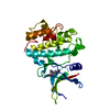
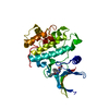
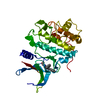
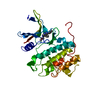
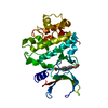
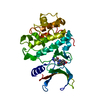
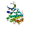
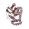
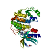
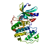
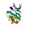
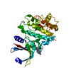
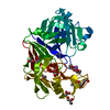
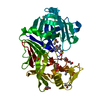
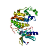
- PDB-2wmt: Crystal structure of checkpoint kinase 1 (Chk1) in complex with i... -
+ Open data

- Basic information
| Entry | Database: PDB / ID: 2wmt | ||||||
|---|---|---|---|---|---|---|---|
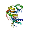
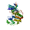
| Title | Crystal structure of checkpoint kinase 1 (Chk1) in complex with inhibitors | ||||||
 Components Components | SERINE/THREONINE-PROTEIN KINASE CHK1 | ||||||
 Keywords Keywords | TRANSFERASE / SERINE/THREONINE-PROTEIN KINASE / POLYMORPHISM / PHOSPHOPROTEIN / UBL CONJUGATION / ISOPEPTIDE BOND / CHECKPOINT KINASE / NUCLEOTIDE-BINDING / SERINE/THREONINE KINASE / DNA DAMAGE / DNA REPAIR / ATP-BINDING / CHK1 / KINASE / NUCLEUS / CYTOPLASM / CELL CYCLE | ||||||
| Function / homology |  Function and homology information Function and homology informationnegative regulation of mitotic nuclear division / apoptotic process involved in development / negative regulation of G0 to G1 transition / histone H3T11 kinase activity / regulation of mitotic centrosome separation / mitotic G2/M transition checkpoint / peptidyl-threonine phosphorylation / inner cell mass cell proliferation / regulation of double-strand break repair via homologous recombination / nucleus organization ...negative regulation of mitotic nuclear division / apoptotic process involved in development / negative regulation of G0 to G1 transition / histone H3T11 kinase activity / regulation of mitotic centrosome separation / mitotic G2/M transition checkpoint / peptidyl-threonine phosphorylation / inner cell mass cell proliferation / regulation of double-strand break repair via homologous recombination / nucleus organization / negative regulation of gene expression, epigenetic / mitotic G2 DNA damage checkpoint signaling / Transcriptional Regulation by E2F6 / replicative senescence / Presynaptic phase of homologous DNA pairing and strand exchange / Activation of ATR in response to replication stress / Chk1/Chk2(Cds1) mediated inactivation of Cyclin B:Cdk1 complex / signal transduction in response to DNA damage / positive regulation of cell cycle / DNA damage checkpoint signaling / regulation of signal transduction by p53 class mediator / replication fork / condensed nuclear chromosome / TP53 Regulates Transcription of DNA Repair Genes / cellular response to mechanical stimulus / Signaling by SCF-KIT / G2/M DNA damage checkpoint / Ubiquitin-Mediated Degradation of Phosphorylated Cdc25A / G2/M transition of mitotic cell cycle / regulation of cell population proliferation / Processing of DNA double-strand break ends / Regulation of TP53 Activity through Phosphorylation / protein phosphorylation / protein kinase activity / DNA replication / non-specific serine/threonine protein kinase / chromatin remodeling / protein domain specific binding / protein serine kinase activity / DNA repair / protein serine/threonine kinase activity / apoptotic process / DNA damage response / centrosome / chromatin / protein-containing complex / : / nucleoplasm / ATP binding / nucleus / cytoplasm / cytosol Similarity search - Function | ||||||
| Biological species |  HOMO SAPIENS (human) HOMO SAPIENS (human) | ||||||
| Method |  X-RAY DIFFRACTION / MOLECULAR REPLACEMENT / Resolution: 2.55 Å X-RAY DIFFRACTION / MOLECULAR REPLACEMENT / Resolution: 2.55 Å | ||||||
 Authors Authors | Matthews, T.P. / Klair, S. / Burns, S. / Boxall, K. / Cherry, M. / Fisher, M. / Westwood, I.M. / Walton, M.I. / McHardy, T. / Cheung, K.-M.J. ...Matthews, T.P. / Klair, S. / Burns, S. / Boxall, K. / Cherry, M. / Fisher, M. / Westwood, I.M. / Walton, M.I. / McHardy, T. / Cheung, K.-M.J. / Van Montfort, R. / Williams, D. / Aherne, G.W. / Garrett, M.D. / Reader, J. / Collins, I. | ||||||
 Citation Citation | Journal: J.Med.Chem. / Year: 2009 Title: Identification of Inhibitors of Checkpoint Kinase 1 Through Template Screening. Authors: Matthews, T.P. / Klair, S. / Burns, S. / Boxall, K. / Cherry, M. / Fisher, M. / Westwood, I.M. / Walton, M.I. / Mchardy, T. / Cheung, K.-M.J. / Van Montfort, R. / Williams, D. / Aherne, G.W. ...Authors: Matthews, T.P. / Klair, S. / Burns, S. / Boxall, K. / Cherry, M. / Fisher, M. / Westwood, I.M. / Walton, M.I. / Mchardy, T. / Cheung, K.-M.J. / Van Montfort, R. / Williams, D. / Aherne, G.W. / Garrett, M.D. / Reader, J. / Collins, I. | ||||||
| History |
|
- Structure visualization
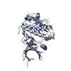
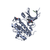
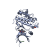
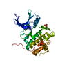
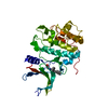
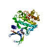
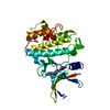
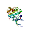
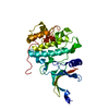
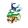
| Structure viewer | Molecule: MolmilJmol/JSmol |
|---|
- Downloads & links
-Download
| PDBx/mmCIF format | 2wmt.cif.gz | 115.4 KB | Display | PDBx/mmCIF format |
|---|---|---|---|---|
| PDB format | pdb2wmt.ent.gz | 88.8 KB | Display | PDB format |
| PDBx/mmJSON format | 2wmt.json.gz | Tree view | PDBx/mmJSON format | |
| Others |  Other downloads Other downloads |
-Validation report
| Arichive directory | https://data.pdbj.org/pub/pdb/validation_reports/wm/2wmtftp://data.pdbj.org/pub/pdb/validation_reports/wm/2wmt | HTTPS FTP |
|---|
-Related structure data
| Related structure data | 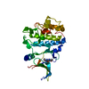 2wmqC 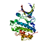 2wmrC 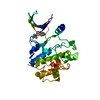 2wmsC 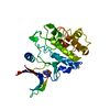 2wmuC 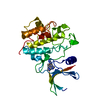 2wmvC 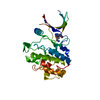 2wmwC 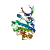 2wmxC  2hy0S C: citing same article ( S: Starting model for refinement |
|---|---|
| Similar structure data |


-Links
 PDBj
PDBj


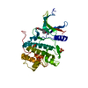
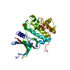
- Assembly
| Deposited unit | 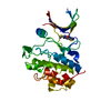
| ||||||||
|---|---|---|---|---|---|---|---|---|---|
| 1 |
| ||||||||
| Unit cell |
|
-Components
| #1: Protein | Mass: 33042.988 Da / Num. of mol.: 1 / Fragment: KINASE DOMAIN, RESIDUES 1-289 Source method: isolated from a genetically manipulated source Source: (gene. exp.) HOMO SAPIENS (human) / Cell line (production host): SF9 / Production host:   SPODOPTERA FRUGIPERDA (fall armyworm) SPODOPTERA FRUGIPERDA (fall armyworm)References: UniProt: O14757, non-specific serine/threonine protein kinase |
|---|---|
| #2: Chemical | ChemComp-ZYT / 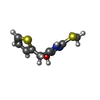  Mass: 226.318 Da / Num. of mol.: 1 / Source method: obtained synthetically / Formula: C9H10N2OS2 Mass: 226.318 Da / Num. of mol.: 1 / Source method: obtained synthetically / Formula: C9H10N2OS2 |
| #3: Water | ChemComp-HOH /  Mass: 18.015 Da / Num. of mol.: 50 / Source method: isolated from a natural source / Formula: H2O Mass: 18.015 Da / Num. of mol.: 50 / Source method: isolated from a natural source / Formula: H2O |
-Experimental details
-Experiment
| Experiment | Method: X-RAY DIFFRACTION |
|---|
- Sample preparation
| Crystal | Density Matthews: 2.7 Å3/Da / Density % sol: 54.1 % / Description: NONE |
|---|---|
| Crystal grow | Details: DL-MALIC ACID/PEG3350 |
-Data collection
| Diffraction | Mean temperature: 100 K |
|---|---|
| Diffraction source | Source: ROTATING ANODE / Type: RIGAKU MICROMAX-007 HF / Wavelength: 1.5418 |
| Detector | Type: RIGAKU CCD / Detector: CCD |
| Radiation | Protocol: SINGLE WAVELENGTH / Monochromatic (M) / Laue (L): M / Scattering type: x-ray |
| Radiation wavelength | Wavelength: 1.5418 Å / Relative weight: 1 |
| Reflection | Resolution: 2.55→32.6 Å / Num. obs: 9862 / % possible obs: 98.3 % / Observed criterion σ(I): 1.5 / Redundancy: 2.5 % / Biso Wilson estimate: 38.54 Å2 / Rmerge(I) obs: 0.08 / Net I/σ(I): 8.8 |
| Reflection shell | Resolution: 2.55→2.69 Å / Redundancy: 2.5 % / Rmerge(I) obs: 0.44 / Mean I/σ(I) obs: 2.5 / % possible all: 99.7 |
- Processing
| Software |
| |||||||||||||||||||||||||||||||||||||||||||||||||||||||||||||||||||||||||||
|---|---|---|---|---|---|---|---|---|---|---|---|---|---|---|---|---|---|---|---|---|---|---|---|---|---|---|---|---|---|---|---|---|---|---|---|---|---|---|---|---|---|---|---|---|---|---|---|---|---|---|---|---|---|---|---|---|---|---|---|---|---|---|---|---|---|---|---|---|---|---|---|---|---|---|---|---|
| Refinement | Method to determine structure: MOLECULAR REPLACEMENT Starting model: PDB ENTRY 2HY0 Resolution: 2.55→32.575 Å / SU ML: 0.38 / σ(F): 0.71 / Phase error: 25.82 / Stereochemistry target values: MLHL
| |||||||||||||||||||||||||||||||||||||||||||||||||||||||||||||||||||||||||||
| Solvent computation | Shrinkage radii: 0.9 Å / VDW probe radii: 1.11 Å / Solvent model: FLAT BULK SOLVENT MODEL / Bsol: 46.454 Å2 / ksol: 0.348 e/Å3 | |||||||||||||||||||||||||||||||||||||||||||||||||||||||||||||||||||||||||||
| Displacement parameters | Biso mean: 46.07 Å2
| |||||||||||||||||||||||||||||||||||||||||||||||||||||||||||||||||||||||||||
| Refinement step | Cycle: LAST / Resolution: 2.55→32.575 Å
| |||||||||||||||||||||||||||||||||||||||||||||||||||||||||||||||||||||||||||
| Refine LS restraints |
| |||||||||||||||||||||||||||||||||||||||||||||||||||||||||||||||||||||||||||
| LS refinement shell |
| |||||||||||||||||||||||||||||||||||||||||||||||||||||||||||||||||||||||||||
| Refinement TLS params. | Method: refined / Refine-ID: X-RAY DIFFRACTION
| |||||||||||||||||||||||||||||||||||||||||||||||||||||||||||||||||||||||||||
| Refinement TLS group |
|
