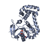
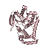
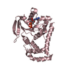
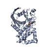
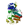
- PDB-3i92: Structure of the cytosolic domain of E. coli FeoB, GppCH2p-bound form -
+
Open data
ID or keywords:
Loading...
-
Basic information
Entry
Database: PDB / ID: 3i92
Title
Structure of the cytosolic domain of E. coli FeoB, GppCH2p-bound form
Components
Ferrous iron transport protein B
Keywords
TRANSPORT PROTEIN / GTPase / GPCR / iron uptake / Feo / Cell inner membrane / Cell membrane / GTP-binding / Ion transport / Iron / Iron transport / Membrane / Nucleotide-binding / Transmembrane / Transport
Function / homology
Function and homology information
iron ion import across plasma membrane / Metal ion assimilation from the host / ferrous iron transmembrane transporter activity / DNA damage response / GTP binding / identical protein binding / plasma membrane Similarity search - Function
Helix Hairpins - #1770 / Ferrous iron transport protein B, C-terminal / Ferrous iron transport protein B C terminus / FeoB, cytosolic helical domain / FeoB cytosolic helical domain / Ferrous iron transport protein B / FeoB-type guanine nucleotide-binding (G) domain / : / Ferrous iron transport protein B / FeoB-type guanine nucleotide-binding (G) domain profile. ...Helix Hairpins - #1770 / Ferrous iron transport protein B, C-terminal / Ferrous iron transport protein B C terminus / FeoB, cytosolic helical domain / FeoB cytosolic helical domain / Ferrous iron transport protein B / FeoB-type guanine nucleotide-binding (G) domain / : / Ferrous iron transport protein B / FeoB-type guanine nucleotide-binding (G) domain profile. / Nucleoside transporter/FeoB GTPase, Gate domain / Nucleoside recognition / Helix Hairpins / P-loop containing nucleotide triphosphate hydrolases / Rossmann fold / P-loop containing nucleoside triphosphate hydrolase / Orthogonal Bundle / 3-Layer(aba) Sandwich / Mainly Alpha / Alpha Beta Similarity search - Domain/homology
Mass: 18.015 Da / Num. of mol.: 16 / Source method: isolated from a natural source / Formula: H2O
-
Experimental details
-
Experiment
Experiment
Method: X-RAY DIFFRACTION / Number of used crystals: 1
-
Sample preparation
Crystal
Density Matthews: 2.1 Å3/Da / Density % sol: 41.53 %
Crystal grow
Temperature: 285 K / Method: vapor diffusion, hanging drop / pH: 7 Details: 30% PEG 3350, 12.5% tacsimate, 250 mM 6-amino hexanoic acid, 100 mM HEPES, pH 7.0, VAPOR DIFFUSION, HANGING DROP, temperature 285K
-
Data collection
Diffraction
Mean temperature: 100 K
Diffraction source
Source: SYNCHROTRON / Site: MAX II / Beamline: I911-2 / Wavelength: 1.04 Å
Detector
Type: MAR CCD 165 mm / Detector: CCD / Date: Nov 11, 2007
Radiation
Monochromator: bent Si (111) crystal, horizontally focusing / Protocol: SINGLE WAVELENGTH / Monochromatic (M) / Laue (L): M / Scattering type: x-ray
Radiation wavelength
Wavelength: 1.04 Å / Relative weight: 1
Reflection
Resolution: 3→45.45 Å / Num. obs: 14810
-
Processing
Software
Name
Version
Classification
ProDC
datacollection
PHASER
phasing
REFMAC
5.3.0037
refinement
MOSFLM
datareduction
SCALA
datascaling
Refinement
Method to determine structure: MOLECULAR REPLACEMENT / Resolution: 3→45.45 Å / Cor.coef. Fo:Fc: 0.896 / Cor.coef. Fo:Fc free: 0.831 / Cross valid method: THROUGHOUT / ESU R Free: 0.553 / Stereochemistry target values: MAXIMUM LIKELIHOOD / Details: HYDROGENS HAVE BEEN ADDED IN THE RIDING POSITIONS
Rfactor
Num. reflection
% reflection
Selection details
Rfree
0.2718
745
5 %
RANDOM
Rwork
0.21658
-
-
-
obs
0.21942
14055
96.62 %
-
all
-
14055
-
-
Solvent computation
Ion probe radii: 0.8 Å / Shrinkage radii: 0.8 Å / VDW probe radii: 1.4 Å / Solvent model: MASK
Displacement parameters
Biso mean: 25.133 Å2
Baniso -1
Baniso -2
Baniso -3
1-
0.02 Å2
0 Å2
-0.01 Å2
2-
-
-0.03 Å2
0 Å2
3-
-
-
0.01 Å2
Refinement step
Cycle: LAST / Resolution: 3→45.45 Å
Protein
Nucleic acid
Ligand
Solvent
Total
Num. atoms
5994
0
99
16
6109
Refine LS restraints
Refine-ID
Type
Dev ideal
Dev ideal target
Number
X-RAY DIFFRACTION
r_bond_refined_d
0.006
0.022
6176
X-RAY DIFFRACTION
r_bond_other_d
0
0.02
4058
X-RAY DIFFRACTION
r_angle_refined_deg
1.015
1.999
8410
X-RAY DIFFRACTION
r_angle_other_deg
4.256
3
9962
X-RAY DIFFRACTION
r_dihedral_angle_1_deg
5.02
5
774
X-RAY DIFFRACTION
r_dihedral_angle_2_deg
34.976
24.778
270
X-RAY DIFFRACTION
r_dihedral_angle_3_deg
16.024
15
1076
X-RAY DIFFRACTION
r_dihedral_angle_4_deg
15.655
15
45
X-RAY DIFFRACTION
r_chiral_restr
0.05
0.2
999
X-RAY DIFFRACTION
r_gen_planes_refined
0.002
0.02
6792
X-RAY DIFFRACTION
r_gen_planes_other
0.002
0.02
1131
X-RAY DIFFRACTION
r_nbd_refined
X-RAY DIFFRACTION
r_nbd_other
X-RAY DIFFRACTION
r_nbtor_refined
X-RAY DIFFRACTION
r_nbtor_other
X-RAY DIFFRACTION
r_xyhbond_nbd_refined
0.127
0.2
133
X-RAY DIFFRACTION
r_xyhbond_nbd_other
X-RAY DIFFRACTION
r_metal_ion_refined
X-RAY DIFFRACTION
r_metal_ion_other
X-RAY DIFFRACTION
r_symmetry_vdw_refined
0.133
0.2
51
X-RAY DIFFRACTION
r_symmetry_vdw_other
0.174
0.2
131
X-RAY DIFFRACTION
r_symmetry_hbond_refined
0.199
0.2
7
X-RAY DIFFRACTION
r_symmetry_hbond_other
X-RAY DIFFRACTION
r_symmetry_metal_ion_refined
X-RAY DIFFRACTION
r_symmetry_metal_ion_other
X-RAY DIFFRACTION
r_mcbond_it
0.199
1.5
3865
X-RAY DIFFRACTION
r_mcbond_other
0
1.5
1574
X-RAY DIFFRACTION
r_mcangle_it
0.364
2
6220
X-RAY DIFFRACTION
r_scbond_it
0.405
3
2311
X-RAY DIFFRACTION
r_scangle_it
0.679
4.5
2190
X-RAY DIFFRACTION
r_rigid_bond_restr
X-RAY DIFFRACTION
r_sphericity_free
X-RAY DIFFRACTION
r_sphericity_bonded
LS refinement shell
Resolution: 3→3.078 Å / Total num. of bins used: 20
Rfactor
Num. reflection
% reflection
Rfree
0.408
45
-
Rwork
0.286
1044
-
obs
-
-
97.84 %
+
About Yorodumi
-
News
-
Feb 9, 2022. New format data for meta-information of EMDB entries
New format data for meta-information of EMDB entries
Version 3 of the EMDB header file is now the official format.
The previous official version 1.9 will be removed from the archive.
In the structure databanks used in Yorodumi, some data are registered as the other names, "COVID-19 virus" and "2019-nCoV". Here are the details of the virus and the list of structure data.
Jan 31, 2019. EMDB accession codes are about to change! (news from PDBe EMDB page)
EMDB accession codes are about to change! (news from PDBe EMDB page)
The allocation of 4 digits for EMDB accession codes will soon come to an end. Whilst these codes will remain in use, new EMDB accession codes will include an additional digit and will expand incrementally as the available range of codes is exhausted. The current 4-digit format prefixed with “EMD-” (i.e. EMD-XXXX) will advance to a 5-digit format (i.e. EMD-XXXXX), and so on. It is currently estimated that the 4-digit codes will be depleted around Spring 2019, at which point the 5-digit format will come into force.
The EM Navigator/Yorodumi systems omit the EMD- prefix.
Related info.:Q: What is EMD? / ID/Accession-code notation in Yorodumi/EM Navigator
Yorodumi is a browser for structure data from EMDB, PDB, SASBDB, etc.
This page is also the successor to EM Navigator detail page, and also detail information page/front-end page for Omokage search.
The word "yorodu" (or yorozu) is an old Japanese word meaning "ten thousand". "mi" (miru) is to see.
Related info.:EMDB / PDB / SASBDB / Comparison of 3 databanks / Yorodumi Search / Aug 31, 2016. New EM Navigator & Yorodumi / Yorodumi Papers / Jmol/JSmol / Function and homology information / Changes in new EM Navigator and Yorodumi
 Movie
Movie Controller

 Yorodumi
 Open data
 Basic information
 Components
 Keywords
 Function and homology information
 X-RAY DIFFRACTION /
  Authors
 Citation
 Structure visualization
 Downloads & links
 Other downloads


 PDBj
PDBj Assembly
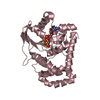

 Mass: 24.305 Da / Num. of mol.: 3 / Source method: obtained synthetically / Formula: Mg
 Mass: 521.208 Da / Num. of mol.: 3 / Source method: obtained synthetically / Formula: C11H18N5O13P3 / Comment: GMP-PCP, energy-carrying molecule analogue*YM
 Mass: 18.015 Da / Num. of mol.: 16 / Source method: isolated from a natural source / Formula: H2O
 Sample preparation
 / Beamline: I911-2 / Wavelength: 1.04 Å
 Processing