 Movie
Movie Controller


+ Open data

- Basic information
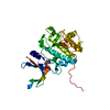
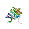
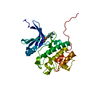
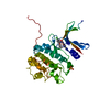
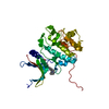
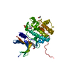
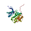
| Entry | Database: PDB / ID: 2hy0 | ||||||
|---|---|---|---|---|---|---|---|
| Title | crystal structure of chek1 in complex with inhibitor 22 | ||||||
 Components Components | Serine/threonine-protein kinase Chk1 | ||||||
 Keywords Keywords | TRANSFERASE / Chek1 / Kinase / Cell cycle checkpoint | ||||||
| Function / homology |  Function and homology information Function and homology informationnegative regulation of mitotic nuclear division / apoptotic process involved in development / negative regulation of G0 to G1 transition / histone H3T11 kinase activity / regulation of mitotic centrosome separation / mitotic G2/M transition checkpoint / inner cell mass cell proliferation / nucleus organization / regulation of double-strand break repair via homologous recombination / peptidyl-threonine phosphorylation ...negative regulation of mitotic nuclear division / apoptotic process involved in development / negative regulation of G0 to G1 transition / histone H3T11 kinase activity / regulation of mitotic centrosome separation / mitotic G2/M transition checkpoint / inner cell mass cell proliferation / nucleus organization / regulation of double-strand break repair via homologous recombination / peptidyl-threonine phosphorylation / mitotic G2 DNA damage checkpoint signaling / negative regulation of gene expression, epigenetic / Transcriptional Regulation by E2F6 / replicative senescence / Presynaptic phase of homologous DNA pairing and strand exchange / Activation of ATR in response to replication stress / Chk1/Chk2(Cds1) mediated inactivation of Cyclin B:Cdk1 complex / signal transduction in response to DNA damage / DNA damage checkpoint signaling / positive regulation of cell cycle / regulation of signal transduction by p53 class mediator / condensed nuclear chromosome / replication fork / TP53 Regulates Transcription of DNA Repair Genes / cellular response to mechanical stimulus / Signaling by SCF-KIT / G2/M DNA damage checkpoint / Ubiquitin-Mediated Degradation of Phosphorylated Cdc25A / G2/M transition of mitotic cell cycle / regulation of cell population proliferation / Processing of DNA double-strand break ends / Regulation of TP53 Activity through Phosphorylation / protein phosphorylation / protein kinase activity / non-specific serine/threonine protein kinase / DNA replication / chromatin remodeling / protein domain specific binding / protein serine kinase activity / DNA repair / protein serine/threonine kinase activity / apoptotic process / centrosome / DNA damage response / chromatin / protein-containing complex / : / nucleoplasm / ATP binding / nucleus / cytosol / cytoplasm Similarity search - Function | ||||||
| Biological species |  Homo sapiens (human) Homo sapiens (human) | ||||||
| Method |  X-RAY DIFFRACTION / FOURIER SYNTHESIS / Resolution: 1.7 Å X-RAY DIFFRACTION / FOURIER SYNTHESIS / Resolution: 1.7 Å | ||||||
 Authors Authors | Yan, Y. | ||||||
 Citation Citation | Journal: Bioorg.Med.Chem.Lett. / Year: 2006 Title: Development of 6-substituted indolylquinolinones as potent Chek1 kinase inhibitors. Authors: Huang, S. / Garbaccio, R.M. / Fraley, M.E. / Steen, J. / Kreatsoulas, C. / Hartman, G. / Stirdivant, S. / Drakas, B. / Rickert, K. / Walsh, E. / Hamilton, K. / Buser, C.A. / Hardwick, J. / ...Authors: Huang, S. / Garbaccio, R.M. / Fraley, M.E. / Steen, J. / Kreatsoulas, C. / Hartman, G. / Stirdivant, S. / Drakas, B. / Rickert, K. / Walsh, E. / Hamilton, K. / Buser, C.A. / Hardwick, J. / Mao, X. / Abrams, M. / Beck, S. / Tao, W. / Lobell, R. / Sepp-Lorenzino, L. / Yan, Y. / Ikuta, M. / Murphy, J.Z. / Sardana, V. / Munshi, S. / Kuo, L. / Reilly, M. / Mahan, E. | ||||||
| History |
|
- Structure visualization
| Structure viewer | Molecule: MolmilJmol/JSmol |
|---|
- Downloads & links
-Download
| PDBx/mmCIF format | 2hy0.cif.gz | 73.5 KB | Display | PDBx/mmCIF format |
|---|---|---|---|---|
| PDB format | pdb2hy0.ent.gz | 53.2 KB | Display | PDB format |
| PDBx/mmJSON format | 2hy0.json.gz | Tree view | PDBx/mmJSON format | |
| Others |  Other downloads Other downloads |
-Validation report
| Arichive directory | https://data.pdbj.org/pub/pdb/validation_reports/hy/2hy0ftp://data.pdbj.org/pub/pdb/validation_reports/hy/2hy0 | HTTPS FTP |
|---|
-Related structure data
| Related structure data |  2hxlC  2hxqC 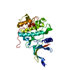 1ia8S C: citing same article ( S: Starting model for refinement |
|---|---|
| Similar structure data |


-Links
 PDBj
PDBj


- Assembly
| Deposited unit | 
| ||||||||
|---|---|---|---|---|---|---|---|---|---|
| 1 |
| ||||||||
| Unit cell |
|
-Components
| #1: Protein | Mass: 36790.957 Da / Num. of mol.: 1 / Fragment: Chek1 kinase domain Source method: isolated from a genetically manipulated source Source: (gene. exp.) Homo sapiens (human) / Gene: CHEK1 / Plasmid: pRAD2030 / Production host:   Spodoptera frugiperda (fall armyworm) Spodoptera frugiperda (fall armyworm)References: UniProt: O14757, non-specific serine/threonine protein kinase |
|---|---|
| #2: Chemical | ChemComp-306 / 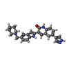  Mass: 423.510 Da / Num. of mol.: 1 / Source method: obtained synthetically / Formula: C26H25N5O Mass: 423.510 Da / Num. of mol.: 1 / Source method: obtained synthetically / Formula: C26H25N5O |
| #3: Water | ChemComp-HOH /  Mass: 18.015 Da / Num. of mol.: 150 / Source method: isolated from a natural source / Formula: H2O Mass: 18.015 Da / Num. of mol.: 150 / Source method: isolated from a natural source / Formula: H2O |
-Experimental details
-Experiment
| Experiment | Method: X-RAY DIFFRACTION / Number of used crystals: 1 |
|---|
- Sample preparation
| Crystal | Density Matthews: 2.31 Å3/Da / Density % sol: 46.78 % |
|---|---|
| Crystal grow | Temperature: 298 K / Method: vapor diffusion, hanging drop / pH: 6.8 Details: 13% PEG 8K, 0.1M ammonium sulfate, 2% glycerol, 0.1M cacodylate buffer at pH 6.8, VAPOR DIFFUSION, HANGING DROP, temperature 298K |
-Data collection
| Diffraction | Mean temperature: 100 K |
|---|---|
| Diffraction source | Source: ROTATING ANODE / Type: RIGAKU / Wavelength: 1.5418 Å |
| Detector | Type: RIGAKU RAXIS IV / Detector: IMAGE PLATE / Date: Dec 6, 2004 / Details: blue confocal optical system |
| Radiation | Protocol: SINGLE WAVELENGTH / Monochromatic (M) / Laue (L): M / Scattering type: x-ray |
| Radiation wavelength | Wavelength: 1.5418 Å / Relative weight: 1 |
| Reflection | Resolution: 1.7→37 Å / Num. all: 37452 / Num. obs: 36749 / % possible obs: 98 % / Observed criterion σ(F): 0 / Observed criterion σ(I): 0 / Redundancy: 4.7 % / Rmerge(I) obs: 0.063 / Rsym value: 0.063 / Net I/σ(I): 10.9 |
| Reflection shell | Resolution: 1.7→1.76 Å / Redundancy: 3.4 % / Rmerge(I) obs: 0.374 / Mean I/σ(I) obs: 3.4 / Num. unique all: 3726 / Rsym value: 0.374 / % possible all: 82 |
- Processing
| Software |
| |||||||||||||||||||||||||
|---|---|---|---|---|---|---|---|---|---|---|---|---|---|---|---|---|---|---|---|---|---|---|---|---|---|---|
| Refinement | Method to determine structure: FOURIER SYNTHESIS Starting model: 1IA8 Resolution: 1.7→37 Å / σ(F): 0 / σ(I): 0 / Stereochemistry target values: Engh & Huber
| |||||||||||||||||||||||||
| Refinement step | Cycle: LAST / Resolution: 1.7→37 Å
| |||||||||||||||||||||||||
| Refine LS restraints |
|
