[English] 日本語
Yorodumi
- PDB-2wm9: Structure of the complex between DOCK9 and Cdc42. -

+
Open data


ID or keywords:

Loading...

-
Basic information

Entry
Database: PDB / ID: 2wm9
TitleStructure of the complex between DOCK9 and Cdc42.
Components
  • CELL DIVISION CONTROL PROTEIN 42 HOMOLOG
  • DEDICATOR OF CYTOKINESIS PROTEIN 9
KeywordsCELL CYCLE / POLYMORPHISM / CELL MEMBRANE / PHOSPHOPROTEIN / NUCLEOTIDE-BINDING / ALTERNATIVE SPLICING / GUANINE-NUCLEOTIDE RELEASING FACTOR / METHYLATION / LIPOPROTEIN / COILED COIL / GTP-BINDING / GEFS / DOCK9 / CDC42 / PRENYLATION
Function / homology
Function and homology information


GBD domain binding / submandibular salivary gland formation / Golgi transport complex / positive regulation of pinocytosis / actin filament branching / positive regulation of synapse structural plasticity / dendritic cell migration / endothelin receptor signaling pathway involved in heart process / cardiac neural crest cell migration involved in outflow tract morphogenesis / storage vacuole ...GBD domain binding / submandibular salivary gland formation / Golgi transport complex / positive regulation of pinocytosis / actin filament branching / positive regulation of synapse structural plasticity / dendritic cell migration / endothelin receptor signaling pathway involved in heart process / cardiac neural crest cell migration involved in outflow tract morphogenesis / storage vacuole / organelle transport along microtubule / positive regulation of epithelial cell proliferation involved in lung morphogenesis / apolipoprotein A-I receptor binding / neuron fate determination / regulation of attachment of spindle microtubules to kinetochore / positive regulation of pseudopodium assembly / GTP-dependent protein binding / Inactivation of CDC42 and RAC1 / cardiac conduction system development / modulation by host of viral process / establishment of Golgi localization / regulation of filopodium assembly / leading edge membrane / neuropilin signaling pathway / positive regulation of intracellular protein transport / regulation of Rho protein signal transduction / cell junction assembly / filopodium assembly / establishment of epithelial cell apical/basal polarity / dendritic spine morphogenesis / mitogen-activated protein kinase kinase kinase binding / thioesterase binding / regulation of modification of postsynaptic structure / regulation of stress fiber assembly / regulation of lamellipodium assembly / adherens junction organization / embryonic heart tube development / RHO GTPases activate KTN1 / DCC mediated attractive signaling / regulation of postsynapse organization / CD28 dependent Vav1 pathway / sprouting angiogenesis / positive regulation of filopodium assembly / Wnt signaling pathway, planar cell polarity pathway / nuclear migration / regulation of mitotic nuclear division / phagocytosis, engulfment / RHOV GTPase cycle / small GTPase-mediated signal transduction / Myogenesis / heart contraction / establishment of cell polarity / establishment or maintenance of cell polarity / Golgi organization / RHOJ GTPase cycle / positive regulation of cytokinesis / RHOQ GTPase cycle / RHO GTPases activate PAKs / RHOU GTPase cycle / CDC42 GTPase cycle / macrophage differentiation / RHOG GTPase cycle / RAC2 GTPase cycle / RHO GTPases Activate WASPs and WAVEs / RAC3 GTPase cycle / spindle midzone / RHO GTPases activate IQGAPs / negative regulation of protein-containing complex assembly / positive regulation of lamellipodium assembly / GPVI-mediated activation cascade / phagocytic vesicle / positive regulation of stress fiber assembly / EPHB-mediated forward signaling / RAC1 GTPase cycle / positive regulation of substrate adhesion-dependent cell spreading / substantia nigra development / positive regulation of GTPase activity / endomembrane system / Gene and protein expression by JAK-STAT signaling after Interleukin-12 stimulation / guanyl-nucleotide exchange factor activity / secretory granule / small monomeric GTPase / actin filament organization / positive regulation of DNA replication / integrin-mediated signaling pathway / filopodium / regulation of actin cytoskeleton organization / FCGR3A-mediated phagocytosis / positive regulation of JNK cascade / RHO GTPases Activate Formins / EGFR downregulation / MAPK6/MAPK4 signaling / Schaffer collateral - CA1 synapse / Regulation of actin dynamics for phagocytic cup formation / cellular response to type II interferon / VEGFA-VEGFR2 Pathway / cytoplasmic ribonucleoprotein granule / G beta:gamma signalling through CDC42 / endocytosis / mitotic spindle
Similarity search - Function
DOCK DHR2 domain, lobe A / DOCK DHR2 domain, lobe C / Dedicator of cytokinesis D, C2 domain / Dedicator of cytokinesis C/D, N-terminal / Dedicator of cytokinesis C/D, N terminal / Dedicator of cytokinesis / C2 DOCK-type domain / DOCKER domain / Dedicator of cytokinesis, C-terminal, lobe A / Dedicator of cytokinesis, C-terminal, lobe C ...DOCK DHR2 domain, lobe A / DOCK DHR2 domain, lobe C / Dedicator of cytokinesis D, C2 domain / Dedicator of cytokinesis C/D, N-terminal / Dedicator of cytokinesis C/D, N terminal / Dedicator of cytokinesis / C2 DOCK-type domain / DOCKER domain / Dedicator of cytokinesis, C-terminal, lobe A / Dedicator of cytokinesis, C-terminal, lobe C / DOCKER, Lobe A / DOCKER, Lobe B / DOCKER, Lobe C / DHR-2, Lobe A / C2 domain in Dock180 and Zizimin proteins / DHR-2, Lobe C / DHR-2, Lobe B / C2 DOCK-type domain profile. / DOCKER domain profile. / Cdc42 / Small GTPase Rho / small GTPase Rho family profile. / PH domain / C2 domain superfamily / PH domain profile. / Pleckstrin homology domain. / Pleckstrin homology domain / Methane Monooxygenase Hydroxylase; Chain G, domain 1 / Rho (Ras homology) subfamily of Ras-like small GTPases / Ras subfamily of RAS small GTPases / Small GTPase / Ras family / Rab subfamily of small GTPases / Serine Threonine Protein Phosphatase 5, Tetratricopeptide repeat / Small GTP-binding protein domain / Alpha Horseshoe / PH-like domain superfamily / Armadillo-type fold / P-loop containing nucleotide triphosphate hydrolases / Up-down Bundle / Rossmann fold / P-loop containing nucleoside triphosphate hydrolase / 3-Layer(aba) Sandwich / Mainly Alpha / Alpha Beta
Similarity search - Domain/homology
Cell division control protein 42 homolog / Dedicator of cytokinesis protein 9
Similarity search - Component
Biological speciesHOMO SAPIENS (human)
MethodX-RAY DIFFRACTION / SYNCHROTRON / SAD / Resolution: 2.2 Å
AuthorsYang, J. / Roe, S.M. / Barford, D.
CitationJournal: Science / Year: 2009
Title: Activation of Rho Gtpases by Dock Exchange Factors is Mediated by a Nucleotide Sensor.
Authors: Yang, J. / Zhang, Z. / Roe, S.M. / Marshall, C.J. / Barford, D.
History
DepositionJun 30, 2009Deposition site: PDBE / Processing site: PDBE
Revision 1.0Sep 22, 2009Provider: repository / Type: Initial release
Revision 1.1May 8, 2011Group: Version format compliance
Revision 1.2Jul 13, 2011Group: Version format compliance
Revision 1.3May 8, 2019Group: Data collection / Experimental preparation / Other
Category: database_PDB_rev / database_PDB_rev_record ...database_PDB_rev / database_PDB_rev_record / exptl_crystal_grow / pdbx_database_proc / pdbx_database_status
Item: _exptl_crystal_grow.method / _pdbx_database_status.recvd_author_approval
Revision 1.4May 15, 2019Group: Data collection / Experimental preparation / Category: exptl_crystal_grow / Item: _exptl_crystal_grow.temp
Revision 1.5May 8, 2024Group: Data collection / Database references ...Data collection / Database references / Derived calculations / Other
Category: chem_comp_atom / chem_comp_bond ...chem_comp_atom / chem_comp_bond / database_2 / pdbx_database_status / struct_site
Item: _database_2.pdbx_DOI / _database_2.pdbx_database_accession ..._database_2.pdbx_DOI / _database_2.pdbx_database_accession / _pdbx_database_status.status_code_sf / _struct_site.pdbx_auth_asym_id / _struct_site.pdbx_auth_comp_id / _struct_site.pdbx_auth_seq_id
Remark 700 
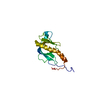
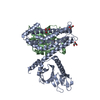
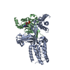
SHEET DETERMINATION METHOD: DSSP THE SHEETS PRESENTED AS "AA" IN EACH CHAIN ON SHEET RECORDS BELOW ... SHEET DETERMINATION METHOD: DSSP THE SHEETS PRESENTED AS "AA" IN EACH CHAIN ON SHEET RECORDS BELOW IS ACTUALLY AN 8-STRANDED BARREL THIS IS REPRESENTED BY A 9-STRANDED SHEET IN WHICH THE FIRST AND LAST STRANDS ARE IDENTICAL.

-
Structure visualization

Structure viewerMolecule:
MolmilJmol/JSmol

Downloads & links

-
Assembly

Deposited unit
A: DEDICATOR OF CYTOKINESIS PROTEIN 9
B: CELL DIVISION CONTROL PROTEIN 42 HOMOLOG
hetero molecules


Theoretical massNumber of molelcules
Total (without water)71,4805
Polymers71,2042
Non-polymers2763
Water3,387188
1
A: DEDICATOR OF CYTOKINESIS PROTEIN 9
B: CELL DIVISION CONTROL PROTEIN 42 HOMOLOG
hetero molecules

A: DEDICATOR OF CYTOKINESIS PROTEIN 9
B: CELL DIVISION CONTROL PROTEIN 42 HOMOLOG
hetero molecules


Theoretical massNumber of molelcules
Total (without water)142,96010
Polymers142,4074
Non-polymers5536
Water724
TypeNameSymmetry operationNumber
identity operation1_555x,y,z1
crystal symmetry operation2_755-x+2,-y,z1
Buried area12430 Å2
ΔGint-67.51 kcal/mol
Surface area49680 Å2
MethodPISA
Unit cell
Length a, b, c (Å)88.410, 93.980, 88.200
Angle α, β, γ (deg.)90.00, 90.00, 90.00
Int Tables number18
Space group name H-MP21212

-
Components

#1: Protein DEDICATOR OF CYTOKINESIS PROTEIN 9 / CDC42 GUANINE NUCLEOTIDE EXCHANGE FACTOR ZIZIMIN-1 / DOCK9


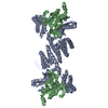
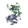
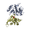
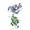
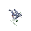
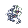
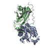
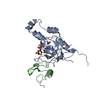
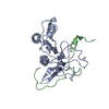
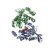
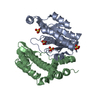
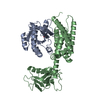
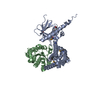
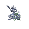
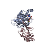
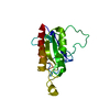
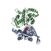
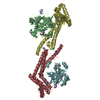
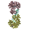
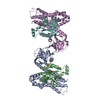
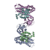
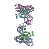
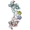
Mass: 50020.320 Da / Num. of mol.: 1 / Fragment: DHR2 DOMAIN, RESIDUES 1605-1652,1676-2053
Source method: isolated from a genetically manipulated source
Source: (gene. exp.) HOMO SAPIENS (human) / Production host: ESCHERICHIA COLI (E. coli) / References: UniProt: Q9BZ29
#2: Protein CELL DIVISION CONTROL PROTEIN 42 HOMOLOG / G25K GTP-BINDING PROTEIN / CDC42


Mass: 21183.357 Da / Num. of mol.: 1
Source method: isolated from a genetically manipulated source
Source: (gene. exp.) HOMO SAPIENS (human) / Production host: ESCHERICHIA COLI (E. coli) / References: UniProt: P60953
#3: Chemical ChemComp-GOL / GLYCEROL / GLYCERIN / PROPANE-1,2,3-TRIOL


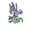
Mass: 92.094 Da / Num. of mol.: 3 / Source method: obtained synthetically / Formula: C3H8O3
#4: Water ChemComp-HOH / water


Mass: 18.015 Da / Num. of mol.: 188 / Source method: isolated from a natural source / Formula: H2O
Sequence detailsISOFORM 2

-
Experimental details

-
Experiment

ExperimentMethod: X-RAY DIFFRACTION / Number of used crystals: 1

-
Sample preparation

CrystalDensity Matthews: 2.33 Å3/Da / Density % sol: 47.2 % / Description: NONE
Crystal growTemperature: 293 K / Method: vapor diffusion, hanging drop / pH: 6
Details: THE PROTEIN WAS CONCENTRATED TO 6 MG/ML. CRYSTALS WERE GROWN USING HANGING DROP VAPOUR DIFFUSION. 1 UL OF COMPLEX WITH 200 MM NDSB-201 ADDED TO PROTEIN PRIOR TO CRYSTALLIZATION WAS MIXED ...Details: THE PROTEIN WAS CONCENTRATED TO 6 MG/ML. CRYSTALS WERE GROWN USING HANGING DROP VAPOUR DIFFUSION. 1 UL OF COMPLEX WITH 200 MM NDSB-201 ADDED TO PROTEIN PRIOR TO CRYSTALLIZATION WAS MIXED WITH EQUAL VOLUME OF CRYSTALLIZATION BUFFER: 14% (W/V) PEG3350,100 MM MES (PH 6.0), 200 MM AMMONIUM ACETATE, 12% GLYCEROL. CRYSTALS WERE GROWN AT 20C.

-
Data collection

DiffractionMean temperature: 100 K
Diffraction sourceSource: SYNCHROTRON / Site: ESRF / Beamline: ID14-2 / Wavelength: 0.933
DetectorType: ADSC CCD / Detector: CCD / Date: Oct 30, 2008
RadiationProtocol: SINGLE WAVELENGTH / Monochromatic (M) / Laue (L): M / Scattering type: x-ray
Radiation wavelengthWavelength: 0.933 Å / Relative weight: 1
ReflectionResolution: 2.2→40 Å / Num. obs: 37540 / % possible obs: 99.1 % / Observed criterion σ(I): 0 / Redundancy: 3.6 % / Biso Wilson estimate: 28.28 Å2 / Rmerge(I) obs: 0.09 / Net I/σ(I): 10.9
Reflection shellResolution: 2.2→2.32 Å / Redundancy: 3.2 % / Rmerge(I) obs: 0.52 / Mean I/σ(I) obs: 2.8 / % possible all: 96.2

-
Processing

Software
NameClassification
iMOSFLMdata reduction
SCALAdata scaling
SHELXphasing
SHARPphasing
PHENIXrefinement
RefinementMethod to determine structure: SAD
Starting model: NONE

Resolution: 2.2→39.463 Å / SU ML: 0.34 / σ(F): 1.17 / Phase error: 25.72 / Stereochemistry target values: ML
RfactorNum. reflection% reflection
Rfree0.2511 3409 5 %
Rwork0.2092 --
obs0.2114 68510 95.08 %
Solvent computationShrinkage radii: 0.9 Å / VDW probe radii: 1.11 Å / Solvent model: FLAT BULK SOLVENT MODEL / Bsol: 46.553 Å2 / ksol: 0.377 e/Å3
Displacement parameters
Baniso -1Baniso -2Baniso -3
1--6.2469 Å20 Å20 Å2
2--0.8127 Å2-0 Å2
3---5.4343 Å2
Refinement stepCycle: LAST / Resolution: 2.2→39.463 Å
ProteinNucleic acidLigandSolventTotal
Num. atoms4585 0 18 188 4791
Refine LS restraints
Refine-IDTypeDev idealNumber
X-RAY DIFFRACTIONf_bond_d0.0124691
X-RAY DIFFRACTIONf_angle_d1.3256354
X-RAY DIFFRACTIONf_dihedral_angle_d17.71688
X-RAY DIFFRACTIONf_chiral_restr0.086722
X-RAY DIFFRACTIONf_plane_restr0.005819
LS refinement shell
Resolution (Å)Rfactor RfreeNum. reflection RfreeRfactor RworkNum. reflection RworkRefine-ID% reflection obs (%)
2.2-2.23150.37871120.32832334X-RAY DIFFRACTION81
2.2315-2.26480.27761240.29692521X-RAY DIFFRACTION88
2.2648-2.30010.30481500.25292674X-RAY DIFFRACTION95
2.3001-2.33790.27751400.24912697X-RAY DIFFRACTION95
2.3379-2.37820.30961290.2532729X-RAY DIFFRACTION95
2.3782-2.42140.26661390.24462665X-RAY DIFFRACTION95
2.4214-2.4680.35551660.25432732X-RAY DIFFRACTION95
2.468-2.51830.29911490.23612708X-RAY DIFFRACTION95
2.5183-2.57310.33351350.22872744X-RAY DIFFRACTION96
2.5731-2.63290.25141400.22212750X-RAY DIFFRACTION96
2.6329-2.69880.26781500.20372727X-RAY DIFFRACTION96
2.6988-2.77170.2621440.20912719X-RAY DIFFRACTION96
2.7717-2.85320.27691370.2142732X-RAY DIFFRACTION96
2.8532-2.94530.31051360.22352764X-RAY DIFFRACTION96
2.9453-3.05050.26261600.20622722X-RAY DIFFRACTION96
3.0505-3.17260.25291390.20072754X-RAY DIFFRACTION96
3.1726-3.31690.24161260.19412769X-RAY DIFFRACTION97
3.3169-3.49170.21631570.18412742X-RAY DIFFRACTION97
3.4917-3.71030.21041550.1772762X-RAY DIFFRACTION97
3.7103-3.99660.20211500.18662775X-RAY DIFFRACTION97
3.9966-4.39830.25921520.16782752X-RAY DIFFRACTION97
4.3983-5.03360.21051540.17442761X-RAY DIFFRACTION97
5.0336-6.33760.22371390.1982783X-RAY DIFFRACTION97
6.3376-39.46930.15571260.18112785X-RAY DIFFRACTION97

+
About Yorodumi

-
News

-
Feb 9, 2022. New format data for meta-information of EMDB entries

New format data for meta-information of EMDB entries

  • Version 3 of the EMDB header file is now the official format.
  • The previous official version 1.9 will be removed from the archive.

Related info.:EMDB header

External links:wwPDB to switch to version 3 of the EMDB data model

-
Aug 12, 2020. Covid-19 info

Covid-19 info

URL: https://pdbj.org/emnavi/covid19.php

New page: Covid-19 featured information page in EM Navigator.

Related info.:Covid-19 info / Mar 5, 2020. Novel coronavirus structure data

+
Mar 5, 2020. Novel coronavirus structure data

Novel coronavirus structure data

Related info.:Yorodumi Speices / Aug 12, 2020. Covid-19 info

External links:COVID-19 featured content - PDBj / Molecule of the Month (242):Coronavirus Proteases

+
Jan 31, 2019. EMDB accession codes are about to change! (news from PDBe EMDB page)

EMDB accession codes are about to change! (news from PDBe EMDB page)

  • The allocation of 4 digits for EMDB accession codes will soon come to an end. Whilst these codes will remain in use, new EMDB accession codes will include an additional digit and will expand incrementally as the available range of codes is exhausted. The current 4-digit format prefixed with “EMD-” (i.e. EMD-XXXX) will advance to a 5-digit format (i.e. EMD-XXXXX), and so on. It is currently estimated that the 4-digit codes will be depleted around Spring 2019, at which point the 5-digit format will come into force.
  • The EM Navigator/Yorodumi systems omit the EMD- prefix.

Related info.:Q: What is EMD? / ID/Accession-code notation in Yorodumi/EM Navigator

External links:EMDB Accession Codes are Changing Soon! / Contact to PDBj

+
Jul 12, 2017. Major update of PDB

Major update of PDB

  • wwPDB released updated PDB data conforming to the new PDBx/mmCIF dictionary.
  • This is a major update changing the version number from 4 to 5, and with Remediation, in which all the entries are updated.
  • In this update, many items about electron microscopy experimental information are reorganized (e.g. em_software).
  • Now, EM Navigator and Yorodumi are based on the updated data.

External links:wwPDB Remediation / Enriched Model Files Conforming to OneDep Data Standards Now Available in the PDB FTP Archive

-
Yorodumi

Thousand views of thousand structures

  • Yorodumi is a browser for structure data from EMDB, PDB, SASBDB, etc.
  • This page is also the successor to EM Navigator detail page, and also detail information page/front-end page for Omokage search.
  • The word "yorodu" (or yorozu) is an old Japanese word meaning "ten thousand". "mi" (miru) is to see.

Related info.:EMDB / PDB / SASBDB / Comparison of 3 databanks / Yorodumi Search / Aug 31, 2016. New EM Navigator & Yorodumi / Yorodumi Papers / Jmol/JSmol / Function and homology information / Changes in new EM Navigator and Yorodumi

Read more