 Movie
Movie Controller


+ Open data

- Basic information
| Entry | Database: PDB / ID: 1a4r | ||||||
|---|---|---|---|---|---|---|---|
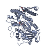
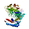
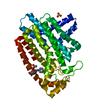
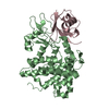
| Title | G12V MUTANT OF HUMAN PLACENTAL CDC42 GTPASE IN THE GDP FORM | ||||||
 Components Components | G25K GTP-BINDING PROTEIN | ||||||
 Keywords Keywords | HYDROLASE / GTPASE / SIGNAL TRANSDUCTION | ||||||
| Function / homology |  Function and homology information Function and homology informationGBD domain binding / positive regulation of pinocytosis / COG complex / endothelin receptor signaling pathway involved in heart process / cardiac neural crest cell migration involved in outflow tract morphogenesis / storage vacuole / dendritic cell migration / neuron fate determination / apolipoprotein A-I receptor binding / positive regulation of epithelial cell proliferation involved in lung morphogenesis ...GBD domain binding / positive regulation of pinocytosis / COG complex / endothelin receptor signaling pathway involved in heart process / cardiac neural crest cell migration involved in outflow tract morphogenesis / storage vacuole / dendritic cell migration / neuron fate determination / apolipoprotein A-I receptor binding / positive regulation of epithelial cell proliferation involved in lung morphogenesis / regulation of attachment of spindle microtubules to kinetochore / organelle transport along microtubule / positive regulation of pseudopodium assembly / Inactivation of CDC42 and RAC1 / cardiac conduction system development / host-mediated perturbation of viral process / leading edge membrane / regulation of filopodium assembly / neuropilin signaling pathway / establishment of Golgi localization / filopodium assembly / cell junction assembly / dendritic spine morphogenesis / establishment of epithelial cell apical/basal polarity / adherens junction organization / GTP-dependent protein binding / thioesterase binding / regulation of lamellipodium assembly / regulation of stress fiber assembly / embryonic heart tube development / RHO GTPases activate KTN1 / DCC mediated attractive signaling / CD28 dependent Vav1 pathway / regulation of postsynapse organization / positive regulation of filopodium assembly / Wnt signaling pathway, planar cell polarity pathway / phagocytosis, engulfment / RHOV GTPase cycle / Myogenesis / nuclear migration / regulation of mitotic nuclear division / small GTPase-mediated signal transduction / positive regulation of cytokinesis / spindle midzone / RHOJ GTPase cycle / heart contraction / RHOQ GTPase cycle / establishment of cell polarity / Golgi organization / RHOU GTPase cycle / establishment or maintenance of cell polarity / RHO GTPases activate PAKs / CDC42 GTPase cycle / macrophage differentiation / RHOG GTPase cycle / RAC3 GTPase cycle / RAC2 GTPase cycle / RHO GTPases Activate WASPs and WAVEs / negative regulation of protein-containing complex assembly / RHO GTPases activate IQGAPs / GPVI-mediated activation cascade / positive regulation of lamellipodium assembly / phagocytic vesicle / positive regulation of stress fiber assembly / RAC1 GTPase cycle / EPHB-mediated forward signaling / positive regulation of substrate adhesion-dependent cell spreading / substantia nigra development / Gene and protein expression by JAK-STAT signaling after Interleukin-12 stimulation / actin filament organization / integrin-mediated signaling pathway / small monomeric GTPase / regulation of actin cytoskeleton organization / FCGR3A-mediated phagocytosis / filopodium / EGFR downregulation / RHO GTPases Activate Formins / Regulation of actin dynamics for phagocytic cup formation / MAPK6/MAPK4 signaling / VEGFA-VEGFR2 Pathway / cellular response to type II interferon / endocytosis / apical part of cell / cytoplasmic ribonucleoprotein granule / cell-cell junction / mitotic spindle / G beta:gamma signalling through CDC42 / intracellular protein localization / ubiquitin protein ligase activity / Factors involved in megakaryocyte development and platelet production / positive regulation of cell growth / actin cytoskeleton organization / G protein activity / midbody / positive regulation of MAPK cascade / positive regulation of phosphatidylinositol 3-kinase/protein kinase B signal transduction / neuron projection / postsynapse / positive regulation of cell migration / Golgi membrane Similarity search - Function | ||||||
| Biological species |  Homo sapiens (human) Homo sapiens (human) | ||||||
| Method |  X-RAY DIFFRACTION / SYNCHROTRON / MOLECULAR REPLACEMENT / Resolution: 2.5 Å X-RAY DIFFRACTION / SYNCHROTRON / MOLECULAR REPLACEMENT / Resolution: 2.5 Å | ||||||
 Authors Authors | Rudolph, M.G. / Vetter, I.R. / Wittinghofer, A. | ||||||
 Citation Citation | Journal: Protein Sci. / Year: 1999 Title: Nucleotide binding to the G12V-mutant of Cdc42 investigated by X-ray diffraction and fluorescence spectroscopy: two different nucleotide states in one crystal. Authors: Rudolph, M.G. / Wittinghofer, A. / Vetter, I.R. | ||||||
| History |
|
- Structure visualization
| Structure viewer | Molecule: MolmilJmol/JSmol |
|---|
- Downloads & links
-Download
| PDBx/mmCIF format | 1a4r.cif.gz | 90.8 KB | Display | PDBx/mmCIF format |
|---|---|---|---|---|
| PDB format | pdb1a4r.ent.gz | 68.4 KB | Display | PDB format |
| PDBx/mmJSON format | 1a4r.json.gz | Tree view | PDBx/mmJSON format | |
| Others |  Other downloads Other downloads |
-Validation report
| Arichive directory | https://data.pdbj.org/pub/pdb/validation_reports/a4/1a4rftp://data.pdbj.org/pub/pdb/validation_reports/a4/1a4r | HTTPS FTP |
|---|
-Related structure data
| Related structure data | 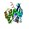 4q21S S: Starting model for refinement |
|---|---|
| Similar structure data |


-Links
 PDBj
PDBj


- Assembly
| Deposited unit | 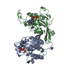
| ||||||||
|---|---|---|---|---|---|---|---|---|---|
| 1 |
| ||||||||
| Unit cell |
| ||||||||
| Noncrystallographic symmetry (NCS) | NCS oper: (Code: given Matrix: (0.5633, 0.8245, -0.0532), Vector: |
-Components
| #1: Protein | Mass: 21325.660 Da / Num. of mol.: 2 / Mutation: YES Source method: isolated from a genetically manipulated source Source: (gene. exp.) Homo sapiens (human) / Tissue: PLACENTA / Cell line: BL21 / Cellular location: CYTOPLASM, INNER SIDE OF MEMBRANE / Plasmid: PGEX2T / Species (production host): Escherichia coli / Cellular location (production host): CYTOPLASM / Production host:  #2: Chemical | ChemComp-MG / |   Mass: 24.305 Da / Num. of mol.: 1 / Source method: obtained synthetically / Formula: Mg Mass: 24.305 Da / Num. of mol.: 1 / Source method: obtained synthetically / Formula: Mg#3: Chemical | ChemComp-GDP / |   Type: RNA linking / Mass: 443.201 Da / Num. of mol.: 1 / Source method: obtained synthetically / Formula: C10H15N5O11P2 / Comment: GDP, energy-carrying molecule*YM Type: RNA linking / Mass: 443.201 Da / Num. of mol.: 1 / Source method: obtained synthetically / Formula: C10H15N5O11P2 / Comment: GDP, energy-carrying molecule*YM#4: Chemical | ChemComp-GNH / |   Mass: 442.216 Da / Num. of mol.: 1 / Source method: obtained synthetically / Formula: C10H16N6O10P2 Mass: 442.216 Da / Num. of mol.: 1 / Source method: obtained synthetically / Formula: C10H16N6O10P2#5: Water | ChemComp-HOH / |  Mass: 18.015 Da / Num. of mol.: 54 / Source method: isolated from a natural source / Formula: H2O Mass: 18.015 Da / Num. of mol.: 54 / Source method: isolated from a natural source / Formula: H2OHas protein modification | Y | |
|---|
-Experimental details
-Experiment
| Experiment | Method: X-RAY DIFFRACTION / Number of used crystals: 1 |
|---|
- Sample preparation
| Crystal | Density Matthews: 2.9 Å3/Da / Density % sol: 51 % | ||||||||||||||||||||||||||||||||||||||||||
|---|---|---|---|---|---|---|---|---|---|---|---|---|---|---|---|---|---|---|---|---|---|---|---|---|---|---|---|---|---|---|---|---|---|---|---|---|---|---|---|---|---|---|---|
| Crystal grow | pH: 4.6 / Details: pH 4.60 | ||||||||||||||||||||||||||||||||||||||||||
| Crystal grow | *PLUS Temperature: 15 ℃ / pH: 7.4 / Method: vapor diffusion, hanging drop | ||||||||||||||||||||||||||||||||||||||||||
| Components of the solutions | *PLUS
|
-Data collection
| Diffraction | Mean temperature: 100 K |
|---|---|
| Diffraction source | Source: SYNCHROTRON / Site: EMBL/DESY, HAMBURG  / Beamline: X31 / Wavelength: 1.0774 / Beamline: X31 / Wavelength: 1.0774 |
| Detector | Type: MARRESEARCH / Detector: IMAGE PLATE / Date: Jun 1, 1997 |
| Radiation | Protocol: SINGLE WAVELENGTH / Monochromatic (M) / Laue (L): M / Scattering type: x-ray |
| Radiation wavelength | Wavelength: 1.0774 Å / Relative weight: 1 |
| Reflection | Resolution: 2.5→30 Å / Num. obs: 17611 / % possible obs: 99.3 % / Observed criterion σ(I): 2 / Redundancy: 4.8 % / Biso Wilson estimate: 46.7 Å2 / Rmerge(I) obs: 0.049 / Net I/σ(I): 15.6 |
| Reflection shell | Resolution: 2.5→2.6 Å / Redundancy: 4.8 % / Rmerge(I) obs: 0.46 / Mean I/σ(I) obs: 3.3 / % possible all: 99.9 |
| Reflection | *PLUS Highest resolution: 2.5 Å / Lowest resolution: 30 Å / Num. obs: 18254 / Observed criterion σ(I): 2 / Redundancy: 4.8 % / Num. measured all: 88071 |
| Reflection shell | *PLUS % possible obs: 99.9 % / Redundancy: 4.8 % / Rmerge(I) obs: 0.46 / Mean I/σ(I) obs: 3.3 |
- Processing
| Software |
| ||||||||||||||||||||||||||||||||||||||||||||||||||||||||||||||||||||||||||||||||
|---|---|---|---|---|---|---|---|---|---|---|---|---|---|---|---|---|---|---|---|---|---|---|---|---|---|---|---|---|---|---|---|---|---|---|---|---|---|---|---|---|---|---|---|---|---|---|---|---|---|---|---|---|---|---|---|---|---|---|---|---|---|---|---|---|---|---|---|---|---|---|---|---|---|---|---|---|---|---|---|---|---|
| Refinement | Method to determine structure: MOLECULAR REPLACEMENT Starting model: 4Q21 WITH ALL NON-CONSERVED RESIDUES MUTATED TO ALANINE Resolution: 2.5→30 Å / Rfactor Rfree error: 0.007 / Data cutoff high absF: 10000000 / Data cutoff low absF: 0.001 / Isotropic thermal model: RESTRAINED / Cross valid method: THROUGHOUT / σ(F): 2 / Details: BULK SOLVENT MODEL USED
| ||||||||||||||||||||||||||||||||||||||||||||||||||||||||||||||||||||||||||||||||
| Displacement parameters | Biso mean: 57.5 Å2 | ||||||||||||||||||||||||||||||||||||||||||||||||||||||||||||||||||||||||||||||||
| Refine analyze |
| ||||||||||||||||||||||||||||||||||||||||||||||||||||||||||||||||||||||||||||||||
| Refinement step | Cycle: LAST / Resolution: 2.5→30 Å
| ||||||||||||||||||||||||||||||||||||||||||||||||||||||||||||||||||||||||||||||||
| Refine LS restraints |
| ||||||||||||||||||||||||||||||||||||||||||||||||||||||||||||||||||||||||||||||||
| Refine LS restraints NCS | NCS model details: CONSTRAINED | ||||||||||||||||||||||||||||||||||||||||||||||||||||||||||||||||||||||||||||||||
| LS refinement shell | Resolution: 2.5→2.54 Å / Rfactor Rfree error: 0.042 / Total num. of bins used: 20
| ||||||||||||||||||||||||||||||||||||||||||||||||||||||||||||||||||||||||||||||||
| Xplor file |
| ||||||||||||||||||||||||||||||||||||||||||||||||||||||||||||||||||||||||||||||||
| Software | *PLUS Name: X-PLOR(ONLINE) / Version: 3.851 / Classification: refinement | ||||||||||||||||||||||||||||||||||||||||||||||||||||||||||||||||||||||||||||||||
| Refinement | *PLUS Rfactor obs: 0.225 / Rfactor Rfree: 0.28 | ||||||||||||||||||||||||||||||||||||||||||||||||||||||||||||||||||||||||||||||||
| Solvent computation | *PLUS | ||||||||||||||||||||||||||||||||||||||||||||||||||||||||||||||||||||||||||||||||
| Displacement parameters | *PLUS | ||||||||||||||||||||||||||||||||||||||||||||||||||||||||||||||||||||||||||||||||
| Refine LS restraints | *PLUS
| ||||||||||||||||||||||||||||||||||||||||||||||||||||||||||||||||||||||||||||||||
| LS refinement shell | *PLUS Rfactor Rfree: 0.362 / Rfactor Rwork: 0.38 |
