 Movie
Movie Controller

[English] 日本語
 Yorodumi
- PDB-1nf3: Structure of Cdc42 in a complex with the GTPase-binding domain of... -
+ Open data

- Basic information
| Entry | Database: PDB / ID: 1nf3 | ||||||
|---|---|---|---|---|---|---|---|
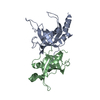
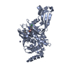
| Title | Structure of Cdc42 in a complex with the GTPase-binding domain of the cell polarity protein, Par6 | ||||||
 Components Components |
| ||||||
 Keywords Keywords | SIGNALING PROTEIN / semi-CRIB motif / Switch I and II / PDZ domain / GTPase binding domain | ||||||
| Function / homology |  Function and homology information Function and homology informationTight junction interactions / RHOV GTPase cycle / regulation of cellular localization / GBD domain binding / positive regulation of pinocytosis / COG complex / endothelin receptor signaling pathway involved in heart process / storage vacuole / cardiac neural crest cell migration involved in outflow tract morphogenesis / dendritic cell migration ...Tight junction interactions / RHOV GTPase cycle / regulation of cellular localization / GBD domain binding / positive regulation of pinocytosis / COG complex / endothelin receptor signaling pathway involved in heart process / storage vacuole / cardiac neural crest cell migration involved in outflow tract morphogenesis / dendritic cell migration / neuron fate determination / apolipoprotein A-I receptor binding / positive regulation of epithelial cell proliferation involved in lung morphogenesis / regulation of attachment of spindle microtubules to kinetochore / organelle transport along microtubule / Inactivation of CDC42 and RAC1 / positive regulation of pseudopodium assembly / cardiac conduction system development / host-mediated perturbation of viral process / leading edge membrane / regulation of filopodium assembly / neuropilin signaling pathway / establishment of Golgi localization / embryonic heart tube development / dendritic spine morphogenesis / filopodium assembly / cell junction assembly / establishment of epithelial cell apical/basal polarity / adherens junction organization / GTP-dependent protein binding / thioesterase binding / regulation of lamellipodium assembly / regulation of stress fiber assembly / RHO GTPases activate KTN1 / DCC mediated attractive signaling / CD28 dependent Vav1 pathway / regulation of postsynapse organization / positive regulation of filopodium assembly / Wnt signaling pathway, planar cell polarity pathway / centrosome cycle / phagocytosis, engulfment / RHOV GTPase cycle / Myogenesis / nuclear migration / regulation of mitotic nuclear division / small GTPase-mediated signal transduction / heart contraction / spindle midzone / positive regulation of cytokinesis / establishment of cell polarity / RHOJ GTPase cycle / Golgi organization / RHOQ GTPase cycle / RHOU GTPase cycle / establishment or maintenance of cell polarity / macrophage differentiation / RHO GTPases activate PAKs / CDC42 GTPase cycle / RHOG GTPase cycle / RAC3 GTPase cycle / RAC2 GTPase cycle / bicellular tight junction / RHO GTPases Activate WASPs and WAVEs / RHO GTPases activate IQGAPs / negative regulation of protein-containing complex assembly / GPVI-mediated activation cascade / positive regulation of lamellipodium assembly / phagocytic vesicle / positive regulation of stress fiber assembly / RAC1 GTPase cycle / positive regulation of substrate adhesion-dependent cell spreading / EPHB-mediated forward signaling / substantia nigra development / Gene and protein expression by JAK-STAT signaling after Interleukin-12 stimulation / integrin-mediated signaling pathway / actin filament organization / regulation of actin cytoskeleton organization / small monomeric GTPase / FCGR3A-mediated phagocytosis / filopodium / EGFR downregulation / RHO GTPases Activate Formins / Regulation of actin dynamics for phagocytic cup formation / cellular response to type II interferon / VEGFA-VEGFR2 Pathway / MAPK6/MAPK4 signaling / endocytosis / apical part of cell / cell junction / cytoplasmic ribonucleoprotein granule / cell-cell junction / mitotic spindle / G beta:gamma signalling through CDC42 / intracellular protein localization / ubiquitin protein ligase activity / Factors involved in megakaryocyte development and platelet production / positive regulation of cell growth / actin cytoskeleton organization / protein-containing complex assembly / G protein activity Similarity search - Function | ||||||
| Biological species |  Homo sapiens (human) Homo sapiens (human) | ||||||
| Method |  X-RAY DIFFRACTION / SYNCHROTRON / MOLECULAR REPLACEMENT / Resolution: 2.1 Å X-RAY DIFFRACTION / SYNCHROTRON / MOLECULAR REPLACEMENT / Resolution: 2.1 Å | ||||||
 Authors Authors | Garrard, S.M. / Capaldo, C.T. / Gao, L. / Rosen, M.K. / Macara, I.G. / Tomchick, D.R. | ||||||
 Citation Citation | Journal: Embo J. / Year: 2003 Title: Structure of Cdc42 in a complex with the GTPase-binding domain of the cell polarity protein, Par6 Authors: Garrard, S.M. / Capaldo, C.T. / Gao, L. / Rosen, M.K. / Macara, I.G. / Tomchick, D.R. #1: Journal: Curr.Biol. / Year: 2002Title: Assembly of epithelial tight junctions is negatively regulated by Par6 Authors: Gao, L. / Joberty, G. / Macara, I.G. #2: Journal: Nat.Cell Biol. / Year: 2000Title: The cell-polarity protein Par6 links Par3 and atypical protein kinase C to Cdc42 Authors: Joberty, G. / Petersen, C. / Gao, L. / Macara, I.G. #3: Journal: Nat.Cell Biol. / Year: 2000Title: A mammalian Par-3-Par-6 complex implicated in Cdc42/Rac1 and aPKC signalling and cell polarity Authors: Lin, D. / Edwards, A.S. / Fawcett, J.P. / Mbamalu, G. / Scott, J.D. / Pawson, T. #4: Journal: Curr.Biol. / Year: 2000Title: A human homolog of the C. elegans polarity determinant Par-6 links Rac and Cdc42 to PKCzeta signaling and cell transformation Authors: Qiu, R.G. / Abo, A. / Steven Martin, G. #5: Journal: DEVELOPMENT / Year: 1996Title: Par-6, a gene involved in the establishment of asymmetry in early C. elegans embryos, mediates the asymmetric localization of Par-3 Authors: Watts, J.L. / Etemad-Moghadam, B. / Guo, S. / Boyd, L. / Draper, B.W. / Mello, C.C. / Priess, J.R. / Kemphues, K.J. | ||||||
| History |
|
- Structure visualization
| Structure viewer | Molecule: MolmilJmol/JSmol |
|---|
- Downloads & links
-Download
| PDBx/mmCIF format | 1nf3.cif.gz | 147 KB | Display | PDBx/mmCIF format |
|---|---|---|---|---|
| PDB format | pdb1nf3.ent.gz | 113.5 KB | Display | PDB format |
| PDBx/mmJSON format | 1nf3.json.gz | Tree view | PDBx/mmJSON format | |
| Others |  Other downloads Other downloads |
-Validation report
| Arichive directory | https://data.pdbj.org/pub/pdb/validation_reports/nf/1nf3ftp://data.pdbj.org/pub/pdb/validation_reports/nf/1nf3 | HTTPS FTP |
|---|
-Related structure data
| Related structure data |  2ngrS S: Starting model for refinement |
|---|---|
| Similar structure data |


-Links
 PDBj
PDBj


- Assembly
| Deposited unit | 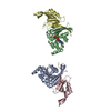
| ||||||||
|---|---|---|---|---|---|---|---|---|---|
| 1 | 
| ||||||||
| 2 | 
| ||||||||
| 3 | 
| ||||||||
| Unit cell |
|
-Components
| #1: Protein | Mass: 21566.949 Da / Num. of mol.: 2 / Mutation: Q61L Source method: isolated from a genetically manipulated source Source: (gene. exp.) Homo sapiens (human) / Gene: CDC42 / Species (production host): Escherichia coli / Production host:  #2: Protein | Mass: 14175.417 Da / Num. of mol.: 2 / Fragment: GTPase-binding domain Source method: isolated from a genetically manipulated source Source: (gene. exp.) #3: Chemical |   Mass: 24.305 Da / Num. of mol.: 2 / Source method: obtained synthetically / Formula: Mg Mass: 24.305 Da / Num. of mol.: 2 / Source method: obtained synthetically / Formula: Mg#4: Chemical |   Mass: 522.196 Da / Num. of mol.: 2 / Source method: obtained synthetically / Formula: C10H17N6O13P3 Mass: 522.196 Da / Num. of mol.: 2 / Source method: obtained synthetically / Formula: C10H17N6O13P3Comment: GppNHp, GMPPNP, energy-carrying molecule analogue*YM #5: Water | ChemComp-HOH / |  Mass: 18.015 Da / Num. of mol.: 423 / Source method: isolated from a natural source / Formula: H2O Mass: 18.015 Da / Num. of mol.: 423 / Source method: isolated from a natural source / Formula: H2OHas protein modification | Y | |
|---|
-Experimental details
-Experiment
| Experiment | Method: X-RAY DIFFRACTION / Number of used crystals: 1 |
|---|
- Sample preparation
| Crystal | Density Matthews: 2.27 Å3/Da / Density % sol: 45.46 % | |||||||||||||||||||||||||||||||||||||||||||||||||||||||||||||||
|---|---|---|---|---|---|---|---|---|---|---|---|---|---|---|---|---|---|---|---|---|---|---|---|---|---|---|---|---|---|---|---|---|---|---|---|---|---|---|---|---|---|---|---|---|---|---|---|---|---|---|---|---|---|---|---|---|---|---|---|---|---|---|---|---|
| Crystal grow | Temperature: 293 K / Method: vapor diffusion, hanging drop / pH: 6.4 Details: 24% PEG 4000, 100mM sodium citrate, 0.2M ammonium acetate, pH 6.4, VAPOR DIFFUSION, HANGING DROP, temperature 293K | |||||||||||||||||||||||||||||||||||||||||||||||||||||||||||||||
| Crystal grow | *PLUS Temperature: 20 ℃ / pH: 8.5 | |||||||||||||||||||||||||||||||||||||||||||||||||||||||||||||||
| Components of the solutions | *PLUS
|
-Data collection
| Diffraction | Mean temperature: 100 K |
|---|---|
| Diffraction source | Source: SYNCHROTRON / Site: APS  / Beamline: 19-ID / Wavelength: 0.9184 Å / Beamline: 19-ID / Wavelength: 0.9184 Å |
| Detector | Type: SBC-2 / Detector: CCD / Date: Mar 8, 2002 / Details: double crystal monochromator |
| Radiation | Monochromator: graphite double crystal / Protocol: SINGLE WAVELENGTH / Monochromatic (M) / Laue (L): M / Scattering type: x-ray |
| Radiation wavelength | Wavelength: 0.9184 Å / Relative weight: 1 |
| Reflection | Resolution: 2.1→40.5 Å / Num. all: 38985 / Num. obs: 38985 / % possible obs: 89.6 % / Observed criterion σ(F): 0 / Observed criterion σ(I): 0 / Redundancy: 2 % / Biso Wilson estimate: 14.7 Å2 / Rmerge(I) obs: 0.104 / Net I/σ(I): 9.2 |
| Reflection shell | Resolution: 2.1→2.14 Å / Rmerge(I) obs: 0.5 / Mean I/σ(I) obs: 2 / % possible all: 87.1 |
| Reflection | *PLUS Num. measured all: 80223 |
| Reflection shell | *PLUS % possible obs: 87.1 % / Rmerge(I) obs: 0.5 |
- Processing
| Software |
| ||||||||||||||||||||||||||||||||||||
|---|---|---|---|---|---|---|---|---|---|---|---|---|---|---|---|---|---|---|---|---|---|---|---|---|---|---|---|---|---|---|---|---|---|---|---|---|---|
| Refinement | Method to determine structure: MOLECULAR REPLACEMENT Starting model: PDB ENTRY 2NGR Resolution: 2.1→29.17 Å / Rfactor Rfree error: 0.007 / Isotropic thermal model: RESTRAINED / Cross valid method: THROUGHOUT / σ(F): 0 / Stereochemistry target values: Engh & Huber
| ||||||||||||||||||||||||||||||||||||
| Solvent computation | Solvent model: FLAT MODEL / Bsol: 46.2756 Å2 / ksol: 0.35546 e/Å3 | ||||||||||||||||||||||||||||||||||||
| Displacement parameters | Biso mean: 28.9 Å2
| ||||||||||||||||||||||||||||||||||||
| Refine analyze |
| ||||||||||||||||||||||||||||||||||||
| Refinement step | Cycle: LAST / Resolution: 2.1→29.17 Å
| ||||||||||||||||||||||||||||||||||||
| Refine LS restraints |
| ||||||||||||||||||||||||||||||||||||
| LS refinement shell | Resolution: 2.1→2.18 Å / Rfactor Rfree error: 0.024 / Total num. of bins used: 10
| ||||||||||||||||||||||||||||||||||||
| Xplor file |
| ||||||||||||||||||||||||||||||||||||
| Refinement | *PLUS Lowest resolution: 30 Å / % reflection Rfree: 5 % | ||||||||||||||||||||||||||||||||||||
| Solvent computation | *PLUS | ||||||||||||||||||||||||||||||||||||
| Displacement parameters | *PLUS | ||||||||||||||||||||||||||||||||||||
| Refine LS restraints | *PLUS
|
