 Movie
Movie Controller

[English] 日本語
 Yorodumi
- PDB-1ees: SOLUTION STRUCTURE OF CDC42HS COMPLEXED WITH A PEPTIDE DERIVED FR... -
+ Open data

- Basic information
| Entry | Database: PDB / ID: 1ees | ||||||
|---|---|---|---|---|---|---|---|
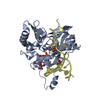
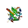
| Title | SOLUTION STRUCTURE OF CDC42HS COMPLEXED WITH A PEPTIDE DERIVED FROM P-21 ACTIVATED KINASE, NMR, 20 STRUCTURES | ||||||
 Components Components |
| ||||||
 Keywords Keywords | STRUCTURAL PROTEIN / protein-protein complex | ||||||
| Function / homology |  Function and homology information Function and homology informationCD28 dependent Vav1 pathway / Ephrin signaling / RHOU GTPase cycle / RHO GTPases activate PAKs / Sema3A PAK dependent Axon repulsion / Generation of second messenger molecules / GBD domain binding / positive regulation of pinocytosis / COG complex / VEGFR2 mediated vascular permeability ...CD28 dependent Vav1 pathway / Ephrin signaling / RHOU GTPase cycle / RHO GTPases activate PAKs / Sema3A PAK dependent Axon repulsion / Generation of second messenger molecules / GBD domain binding / positive regulation of pinocytosis / COG complex / VEGFR2 mediated vascular permeability / RAC1 GTPase cycle / endothelin receptor signaling pathway involved in heart process / storage vacuole / cardiac neural crest cell migration involved in outflow tract morphogenesis / dendritic cell migration / neuron fate determination / apolipoprotein A-I receptor binding / positive regulation of epithelial cell proliferation involved in lung morphogenesis / regulation of attachment of spindle microtubules to kinetochore / organelle transport along microtubule / positive regulation of pseudopodium assembly / dendritic spine development / MAPK6/MAPK4 signaling / Inactivation of CDC42 and RAC1 / CD209 (DC-SIGN) signaling / cardiac conduction system development / host-mediated perturbation of viral process / leading edge membrane / regulation of filopodium assembly / neuropilin signaling pathway / regulation of actin filament polymerization / establishment of Golgi localization / filopodium assembly / positive regulation of dendritic spine morphogenesis / cell junction assembly / dendritic spine morphogenesis / establishment of epithelial cell apical/basal polarity / adherens junction organization / GTP-dependent protein binding / thioesterase binding / regulation of lamellipodium assembly / embryonic heart tube development / regulation of stress fiber assembly / RHO GTPases activate KTN1 / DCC mediated attractive signaling / CD28 dependent Vav1 pathway / regulation of postsynapse organization / positive regulation of fibroblast migration / positive regulation of filopodium assembly / Wnt signaling pathway, planar cell polarity pathway / phagocytosis, engulfment / positive regulation of DNA biosynthetic process / RHOV GTPase cycle / Myogenesis / nuclear migration / regulation of mitotic nuclear division / small GTPase-mediated signal transduction / regulation of axonogenesis / heart contraction / regulation of neuron projection development / spindle midzone / positive regulation of cytokinesis / RHOJ GTPase cycle / RHOQ GTPase cycle / Golgi organization / establishment of cell polarity / MAP kinase kinase activity / dendrite development / RHOU GTPase cycle / establishment or maintenance of cell polarity / CDC42 GTPase cycle / RHO GTPases activate PAKs / macrophage differentiation / regulation of MAPK cascade / RHOG GTPase cycle / RAC3 GTPase cycle / RAC2 GTPase cycle / RHO GTPases Activate WASPs and WAVEs / negative regulation of protein-containing complex assembly / RHO GTPases activate IQGAPs / GPVI-mediated activation cascade / positive regulation of lamellipodium assembly / phagocytic vesicle / positive regulation of stress fiber assembly / RAC1 GTPase cycle / EPHB-mediated forward signaling / positive regulation of substrate adhesion-dependent cell spreading / axonogenesis / substantia nigra development / Gene and protein expression by JAK-STAT signaling after Interleukin-12 stimulation / cellular response to starvation / integrin-mediated signaling pathway / actin filament organization / small monomeric GTPase / regulation of actin cytoskeleton organization / FCGR3A-mediated phagocytosis / filopodium / EGFR downregulation / RHO GTPases Activate Formins / SH3 domain binding Similarity search - Function | ||||||
| Biological species |  Homo sapiens (human) Homo sapiens (human) | ||||||
| Method | SOLUTION NMR / distance geometry simulated annealing Ramachandran refinement | ||||||
 Authors Authors | Gizachew, D. / Guo, W. / Chohan, K.C. / Sutcliffe, M.J. / Oswald, R.E. | ||||||
 Citation Citation | Journal: Biochemistry / Year: 2000 Title: Structure of the complex of Cdc42Hs with a peptide derived from P-21 activated kinase. Authors: Gizachew, D. / Guo, W. / Chohan, K.K. / Sutcliffe, M.J. / Oswald, R.E. #1: Journal: Biochemistry / Year: 1999Title: Backbone Dynamics of Inactive, Active, and Effector-Bound Cdc42Hs from Measurements of (15)N Relaxation Parameters at Multiple Field Strengths Authors: Loh, A.P. / Guo, W. / Nicholson, L.K. / Oswald, R.E. #2: Journal: Biochemistry / Year: 1998Title: Identification of the Binding Surface on Cdc42Hs for p21-Activated Kinase Authors: Guo, W. / Sutcliffe, M.J. / Cerione, R.A. / Oswald, R.E. #3: Journal: Biochemistry / Year: 1997Title: Definition of the Switch Surface in the Solution Structure of Cdc42Hs Authors: Feltham, J.L. / Dotsch, V. / Raza, S. / Manor, D. / Cerione, R.A. / Sutcliffe, M.J. / Wagner, G. / Oswald, R.E. | ||||||
| History |
|
- Structure visualization
| Structure viewer | Molecule: MolmilJmol/JSmol |
|---|
- Downloads & links
-Download
| PDBx/mmCIF format | 1ees.cif.gz | 1.3 MB | Display | PDBx/mmCIF format |
|---|---|---|---|---|
| PDB format | pdb1ees.ent.gz | 1.1 MB | Display | PDB format |
| PDBx/mmJSON format | 1ees.json.gz | Tree view | PDBx/mmJSON format | |
| Others |  Other downloads Other downloads |
-Validation report
| Arichive directory | https://data.pdbj.org/pub/pdb/validation_reports/ee/1eesftp://data.pdbj.org/pub/pdb/validation_reports/ee/1ees | HTTPS FTP |
|---|
-Related structure data
| Related structure data | |
|---|---|
| Similar structure data |


-Links
 PDBj
PDBj


- Assembly
| Deposited unit | 
| |||||||||
|---|---|---|---|---|---|---|---|---|---|---|
| 1 |
| |||||||||
| NMR ensembles |
|
-Components
| #1: Protein | Mass: 19774.705 Da / Num. of mol.: 1 / Fragment: AMINO ACIDS 1-178 Source method: isolated from a genetically manipulated source Source: (gene. exp.) Homo sapiens (human) / Organ: PLACENTA / Plasmid: PET-15B / Production host:  |
|---|---|
| #2: Protein/peptide | Mass: 5117.615 Da / Num. of mol.: 1 / Fragment: AMINO ACIDS 65-108 Source method: isolated from a genetically manipulated source Source: (gene. exp.) |
-Experimental details
-Experiment
| Experiment | Method: SOLUTION NMR | ||||||||||||||||||||||||||||||||||||
|---|---|---|---|---|---|---|---|---|---|---|---|---|---|---|---|---|---|---|---|---|---|---|---|---|---|---|---|---|---|---|---|---|---|---|---|---|---|
| NMR experiment |
| ||||||||||||||||||||||||||||||||||||
| NMR details | Text: The structure was determined using triple-resonance and double-resonance NMR spectroscopy |
- Sample preparation
| Details |
| |||||||||||||||||||||||||||
|---|---|---|---|---|---|---|---|---|---|---|---|---|---|---|---|---|---|---|---|---|---|---|---|---|---|---|---|---|
| Sample conditions | Ionic strength: 64 mM / pH: 5.5 / Pressure: ambient / Temperature: 298 K | |||||||||||||||||||||||||||
| Crystal grow | *PLUS Method: other / Details: NMR |
-NMR measurement
| NMR spectrometer | Type: Varian INOVA / Manufacturer: Varian / Model: INOVA / Field strength: 600 MHz |
|---|
- Processing
| NMR software |
| ||||||||||||||||||||||||||||
|---|---|---|---|---|---|---|---|---|---|---|---|---|---|---|---|---|---|---|---|---|---|---|---|---|---|---|---|---|---|
| Refinement | Method: distance geometry simulated annealing Ramachandran refinement Software ordinal: 1 Details: Structures are based on 2412 distance and dihedral restraints | ||||||||||||||||||||||||||||
| NMR representative | Selection criteria: closest to the average | ||||||||||||||||||||||||||||
| NMR ensemble | Conformer selection criteria: structures with acceptable covalent geometry,structures with favorable non-bond energy,structures with the least restraint violations,structures with the lowest energy Conformers calculated total number: 200 / Conformers submitted total number: 20 |
 NMRPipe