 Movie
Movie Controller


+ Open data

- Basic information
| Entry | Database: PDB / ID: 1am4 | ||||||
|---|---|---|---|---|---|---|---|
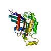
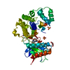
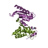
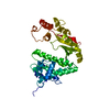
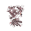
| Title | COMPLEX BETWEEN CDC42HS.GMPPNP AND P50 RHOGAP (H. SAPIENS) | ||||||
 Components Components |
| ||||||
 Keywords Keywords | COMPLEX (GTPASE-ACTIVATING/GTP-BINDING) / COMPLEX (GTPASE-ACTIVATING-GTP-BINDING) / GTPASE ACTIVATION / COMPLEX (GTPASE-ACTIVATING-GTP-BINDING) complex | ||||||
| Function / homology |  Function and homology information Function and homology informationnegative regulation of endocytic recycling / GBD domain binding / positive regulation of pinocytosis / COG complex / storage vacuole / cardiac neural crest cell migration involved in outflow tract morphogenesis / apolipoprotein A-I receptor binding / positive regulation of epithelial cell proliferation involved in lung morphogenesis / regulation of attachment of spindle microtubules to kinetochore / organelle transport along microtubule ...negative regulation of endocytic recycling / GBD domain binding / positive regulation of pinocytosis / COG complex / storage vacuole / cardiac neural crest cell migration involved in outflow tract morphogenesis / apolipoprotein A-I receptor binding / positive regulation of epithelial cell proliferation involved in lung morphogenesis / regulation of attachment of spindle microtubules to kinetochore / organelle transport along microtubule / Inactivation of CDC42 and RAC1 / endothelin receptor signaling pathway / embryonic heart tube development / positive regulation of pseudopodium assembly / host-mediated perturbation of viral process / leading edge membrane / regulation of filopodium assembly / neuropilin signaling pathway / establishment of Golgi localization / dendritic spine morphogenesis / cell junction assembly / establishment of epithelial cell apical/basal polarity / heart process / GTP-dependent protein binding / thioesterase binding / regulation of lamellipodium assembly / regulation of stress fiber assembly / RHOD GTPase cycle / regulation of small GTPase mediated signal transduction / RHO GTPases activate KTN1 / DCC mediated attractive signaling / positive regulation of filopodium assembly / RHOF GTPase cycle / CD28 dependent Vav1 pathway / regulation of postsynapse organization / RND2 GTPase cycle / Wnt signaling pathway, planar cell polarity pathway / phagocytosis, engulfment / endosomal transport / RHOV GTPase cycle / RHOB GTPase cycle / Myogenesis / small GTPase-mediated signal transduction / establishment of cell polarity / positive regulation of cytokinesis / RHOJ GTPase cycle / spindle midzone / RHOC GTPase cycle / Golgi organization / RHOQ GTPase cycle / RHOU GTPase cycle / macrophage differentiation / establishment or maintenance of cell polarity / CDC42 GTPase cycle / RHO GTPases activate PAKs / RHOG GTPase cycle / RAC3 GTPase cycle / RHOA GTPase cycle / RAC2 GTPase cycle / RHO GTPases Activate WASPs and WAVEs / negative regulation of protein-containing complex assembly / positive regulation of lamellipodium assembly / RHO GTPases activate IQGAPs / Rho protein signal transduction / positive regulation of stress fiber assembly / GPVI-mediated activation cascade / ruffle / positive regulation of substrate adhesion-dependent cell spreading / phagocytic vesicle / RAC1 GTPase cycle / EPHB-mediated forward signaling / substantia nigra development / Gene and protein expression by JAK-STAT signaling after Interleukin-12 stimulation / integrin-mediated signaling pathway / actin filament organization / GTPase activator activity / regulation of actin cytoskeleton organization / small monomeric GTPase / FCGR3A-mediated phagocytosis / filopodium / transferrin transport / EGFR downregulation / RHO GTPases Activate Formins / SH3 domain binding / Regulation of actin dynamics for phagocytic cup formation / VEGFA-VEGFR2 Pathway / MAPK6/MAPK4 signaling / small GTPase binding / mitotic spindle / endocytosis / apical part of cell / cytoplasmic ribonucleoprotein granule / cell-cell junction / G beta:gamma signalling through CDC42 / microtubule cytoskeleton / ubiquitin protein ligase activity / Factors involved in megakaryocyte development and platelet production / positive regulation of cell growth / actin cytoskeleton organization / G protein activity Similarity search - Function | ||||||
| Biological species |  Homo sapiens (human) Homo sapiens (human) | ||||||
| Method |  X-RAY DIFFRACTION / SYNCHROTRON / MOLECULAR REPLACEMENT / Resolution: 2.7 Å X-RAY DIFFRACTION / SYNCHROTRON / MOLECULAR REPLACEMENT / Resolution: 2.7 Å | ||||||
 Authors Authors | Rittinger, K. / Walker, P. / Gamblin, S.J. / Smerdon, S.J. | ||||||
 Citation Citation | Journal: Nature / Year: 1997 Title: Crystal structure of a small G protein in complex with the GTPase-activating protein rhoGAP. Authors: Rittinger, K. / Walker, P.A. / Eccleston, J.F. / Nurmahomed, K. / Owen, D. / Laue, E. / Gamblin, S.J. / Smerdon, S.J. | ||||||
| History |
|
- Structure visualization
| Structure viewer | Molecule: MolmilJmol/JSmol |
|---|
- Downloads & links
-Download
| PDBx/mmCIF format | 1am4.cif.gz | 240.3 KB | Display | PDBx/mmCIF format |
|---|---|---|---|---|
| PDB format | pdb1am4.ent.gz | 187.9 KB | Display | PDB format |
| PDBx/mmJSON format | 1am4.json.gz | Tree view | PDBx/mmJSON format | |
| Others |  Other downloads Other downloads |
-Validation report
| Arichive directory | https://data.pdbj.org/pub/pdb/validation_reports/am/1am4ftp://data.pdbj.org/pub/pdb/validation_reports/am/1am4 | HTTPS FTP |
|---|
-Related structure data


-Links
 PDBj
PDBj


- Assembly
| Deposited unit | 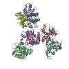
| ||||||||||||
|---|---|---|---|---|---|---|---|---|---|---|---|---|---|
| 1 | 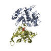
| ||||||||||||
| 2 | 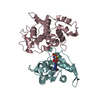
| ||||||||||||
| 3 | 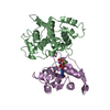
| ||||||||||||
| Unit cell |
| ||||||||||||
| Noncrystallographic symmetry (NCS) | NCS oper:
|
-Components
| #1: Protein | Mass: 22741.998 Da / Num. of mol.: 3 Source method: isolated from a genetically manipulated source Source: (gene. exp.) Homo sapiens (human) / Cellular location: CYTOPLASM / Production host:  #2: Protein | Mass: 19639.521 Da / Num. of mol.: 3 / Mutation: M501P Source method: isolated from a genetically manipulated source Details: HETERODIMER / Source: (gene. exp.) Homo sapiens (human) / Cellular location: CYTOPLASM / Production host: #3: Chemical |   Mass: 24.305 Da / Num. of mol.: 3 / Source method: obtained synthetically / Formula: Mg Mass: 24.305 Da / Num. of mol.: 3 / Source method: obtained synthetically / Formula: Mg#4: Chemical |   Mass: 522.196 Da / Num. of mol.: 3 / Source method: obtained synthetically / Formula: C10H17N6O13P3 Mass: 522.196 Da / Num. of mol.: 3 / Source method: obtained synthetically / Formula: C10H17N6O13P3Comment: GppNHp, GMPPNP, energy-carrying molecule analogue*YM #5: Water | ChemComp-HOH / |  Mass: 18.015 Da / Num. of mol.: 302 / Source method: isolated from a natural source / Formula: H2O Mass: 18.015 Da / Num. of mol.: 302 / Source method: isolated from a natural source / Formula: H2O |
|---|
-Experimental details
-Experiment
| Experiment | Method: X-RAY DIFFRACTION / Number of used crystals: 1 |
|---|
- Sample preparation
| Crystal | Density Matthews: 3.75 Å3/Da / Density % sol: 55 % | ||||||||||||||||||||||||||||||||||||
|---|---|---|---|---|---|---|---|---|---|---|---|---|---|---|---|---|---|---|---|---|---|---|---|---|---|---|---|---|---|---|---|---|---|---|---|---|---|
| Crystal grow | pH: 5.9 / Details: pH 5.9 | ||||||||||||||||||||||||||||||||||||
| Crystal | *PLUS | ||||||||||||||||||||||||||||||||||||
| Crystal grow | *PLUS Temperature: 4 ℃ / Method: vapor diffusion | ||||||||||||||||||||||||||||||||||||
| Components of the solutions | *PLUS
|
-Data collection
| Diffraction | Mean temperature: 100 K |
|---|---|
| Diffraction source | Source: SYNCHROTRON / Site: SRS  / Beamline: PX7.2 / Wavelength: 1.488 / Beamline: PX7.2 / Wavelength: 1.488 |
| Detector | Type: MARRESEARCH / Detector: IMAGE PLATE / Date: Mar 1, 1997 |
| Radiation | Monochromatic (M) / Laue (L): M / Scattering type: x-ray |
| Radiation wavelength | Wavelength: 1.488 Å / Relative weight: 1 |
| Reflection | Resolution: 2.7→25 Å / Num. obs: 49357 / % possible obs: 97.6 % / Redundancy: 5 % / Biso Wilson estimate: 25 Å2 / Rmerge(I) obs: 0.064 / Net I/σ(I): 26 |
| Reflection shell | Resolution: 2.7→2.8 Å / Redundancy: 3.5 % / Rmerge(I) obs: 0.247 / Mean I/σ(I) obs: 5 / % possible all: 96.5 |
| Reflection | *PLUS Num. measured all: 246060 |
| Reflection shell | *PLUS % possible obs: 96.5 % |
- Processing
| Software |
| |||||||||||||||||||||||||||||||||||||||||||||||||||||||||||||||
|---|---|---|---|---|---|---|---|---|---|---|---|---|---|---|---|---|---|---|---|---|---|---|---|---|---|---|---|---|---|---|---|---|---|---|---|---|---|---|---|---|---|---|---|---|---|---|---|---|---|---|---|---|---|---|---|---|---|---|---|---|---|---|---|---|
| Refinement | Method to determine structure: MOLECULAR REPLACEMENT Starting model: PDB ENTRIES 1RGP, 1MH1 Resolution: 2.7→12 Å / Cross valid method: THROUGHOUT
| |||||||||||||||||||||||||||||||||||||||||||||||||||||||||||||||
| Displacement parameters | Biso mean: 18 Å2 | |||||||||||||||||||||||||||||||||||||||||||||||||||||||||||||||
| Refinement step | Cycle: LAST / Resolution: 2.7→12 Å
| |||||||||||||||||||||||||||||||||||||||||||||||||||||||||||||||
| Refine LS restraints |
| |||||||||||||||||||||||||||||||||||||||||||||||||||||||||||||||
| Software | *PLUS Name: CCP4 / Classification: refinement | |||||||||||||||||||||||||||||||||||||||||||||||||||||||||||||||
| Refinement | *PLUS Rfactor obs: 0.231 | |||||||||||||||||||||||||||||||||||||||||||||||||||||||||||||||
| Solvent computation | *PLUS | |||||||||||||||||||||||||||||||||||||||||||||||||||||||||||||||
| Displacement parameters | *PLUS | |||||||||||||||||||||||||||||||||||||||||||||||||||||||||||||||
| Refine LS restraints | *PLUS Type: p_angle_deg / Dev ideal: 2.7 |
