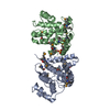
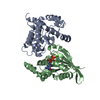
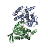
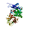
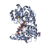
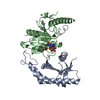
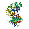
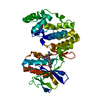
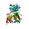
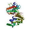
Entry Database : PDB / ID : 5c2kTitle Crystal structure of the fusion protein linked by RhoA and the GAP domain of MgcRacGAP Transforming protein RhoA,Rac GTPase-activating protein 1 Keywords / / / Function / homology Function Domain/homology Component
/ / / / / / / / / / / / / / / / / / / / / / / / / / / / / / / / / / / / / / / / / / / / / / / / / / / / / / / / / / / / / / / / / / / / / / / / / / / / / / / / / / / / / / / / / / / / / / / / / / / / / / / / / / / / / / / / / / / / / / / / / / / / / / / / / / / / / / / / / / / / / / / / Biological species Homo sapiens (human)Method / / / Resolution : 1.42 Å Authors Murayama, K. / Kato-Murayama, M. / Hosaka, T. / Kitamura, T. / Yokoyama, S. / Shirouzu, M. Journal : J.Struct.Biol. / Year : 2024Title : Structural basis for the effects of Ser387 phosphorylation of MgcRacGAP on its GTPase-activating activities for CDC42 and RHOA.Authors : Murayama, K. / Kato-Murayama, M. / Hosaka, T. / Kitamura, T. / Yokoyama, S. / Shirouzu, M. History Deposition Jun 16, 2015 Deposition site / Processing site Revision 1.0 Jun 22, 2016 Provider / Type Revision 1.1 Feb 19, 2020 Group / Derived calculations / Category / pdbx_struct_oper_listItem / _pdbx_struct_oper_list.symmetry_operationRevision 1.2 Nov 8, 2023 Group Data collection / Database references ... Data collection / Database references / Derived calculations / Refinement description Category chem_comp_atom / chem_comp_bond ... chem_comp_atom / chem_comp_bond / database_2 / pdbx_initial_refinement_model / pdbx_struct_conn_angle / struct_conn Item _database_2.pdbx_DOI / _database_2.pdbx_database_accession ... _database_2.pdbx_DOI / _database_2.pdbx_database_accession / _pdbx_struct_conn_angle.ptnr1_auth_seq_id / _pdbx_struct_conn_angle.ptnr3_auth_seq_id / _pdbx_struct_conn_angle.value / _struct_conn.pdbx_dist_value / _struct_conn.ptnr2_auth_seq_id Revision 1.3 Dec 4, 2024 Group / Structure summary / Category / citation_author / pdbx_entry_detailsItem _citation.country / _citation.journal_abbrev ... _citation.country / _citation.journal_abbrev / _citation.journal_id_ASTM / _citation.journal_id_CSD / _citation.journal_id_ISSN / _citation.journal_volume / _citation.page_first / _citation.page_last / _citation.pdbx_database_id_DOI / _citation.pdbx_database_id_PubMed / _citation.title / _citation.year / _pdbx_entry_details.has_protein_modification
Show all Show less
 Movie
Movie Controller

 Yorodumi
 Open data
 Basic information
 Components
 Keywords
 Function and homology information
 Homo sapiens (human)
 X-RAY DIFFRACTION /
  Authors
 Citation
 Structure visualization
 Downloads & links
 Other downloads


 PDBj
PDBj


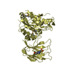
 Assembly

 Mass: 24.305 Da / Num. of mol.: 1 / Source method: obtained synthetically / Formula: Mg
 Mass: 83.977 Da / Num. of mol.: 1 / Source method: obtained synthetically / Formula: AlF3
 Type: RNA linking / Mass: 443.201 Da / Num. of mol.: 1 / Source method: obtained synthetically / Formula: C10H15N5O11P2 / Comment: GDP, energy-carrying molecule*YM
 Mass: 18.015 Da / Num. of mol.: 609 / Source method: isolated from a natural source / Formula: H2O
 Sample preparation
 / Beamline: BL32XU / Wavelength: 1 Å
 Processing