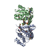
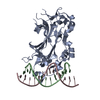
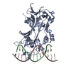
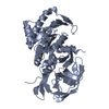
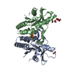
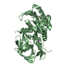
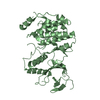
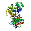
[English] 日本語
Yorodumi
- PDB-5c2j: Complex structure of the GAP domain of MgcRacGAP and Cdc42 -

+
Open data


ID or keywords:

Loading...

-
Basic information

Entry
Database: PDB / ID: 5c2j
TitleComplex structure of the GAP domain of MgcRacGAP and Cdc42
Components
  • Cell division control protein 42 homolog
  • Rac GTPase-activating protein 1
KeywordsHYDROLASE ACTIVATOR/SIGNALING PROTEIN / GTPase activation / Complex / small G-protein / HYDROLASE ACTIVATOR-SIGNALING PROTEIN complex
Function / homology
Function and homology information


DCC mediated attractive signaling / RHO GTPases activate KTN1 / centralspindlin complex / RHOV GTPase cycle / CD28 dependent Vav1 pathway / RHOQ GTPase cycle / RHOU GTPase cycle / RAC3 GTPase cycle / establishment or maintenance of apical/basal cell polarity / RAC2 GTPase cycle ...DCC mediated attractive signaling / RHO GTPases activate KTN1 / centralspindlin complex / RHOV GTPase cycle / CD28 dependent Vav1 pathway / RHOQ GTPase cycle / RHOU GTPase cycle / RAC3 GTPase cycle / establishment or maintenance of apical/basal cell polarity / RAC2 GTPase cycle / RHO GTPases activate PAKs / CDC42 GTPase cycle / actomyosin contractile ring assembly / G beta:gamma signalling through CDC42 / GPVI-mediated activation cascade / EPHB-mediated forward signaling / RHO GTPases Activate WASPs and WAVEs / nucleus localization / EGFR downregulation / GBD domain binding / submandibular salivary gland formation / Golgi transport complex / positive regulation of pinocytosis / actin filament branching / RAC1 GTPase cycle / positive regulation of synapse structural plasticity / dendritic cell migration / endothelin receptor signaling pathway involved in heart process / mitotic spindle midzone assembly / sulfate transmembrane transport / cardiac neural crest cell migration involved in outflow tract morphogenesis / storage vacuole / RHO GTPases activate IQGAPs / organelle transport along microtubule / positive regulation of epithelial cell proliferation involved in lung morphogenesis / apolipoprotein A-I receptor binding / RHO GTPases Activate Formins / neuron fate determination / regulation of attachment of spindle microtubules to kinetochore / RHOG GTPase cycle / positive regulation of pseudopodium assembly / GTP-dependent protein binding / MAPK6/MAPK4 signaling / Regulation of actin dynamics for phagocytic cup formation / cardiac conduction system development / modulation by host of viral process / Factors involved in megakaryocyte development and platelet production / heart process / establishment of Golgi localization / regulation of filopodium assembly / leading edge membrane / neuropilin signaling pathway / positive regulation of intracellular protein transport / Myogenesis / cell junction assembly / filopodium assembly / establishment of epithelial cell apical/basal polarity / dendritic spine morphogenesis / VEGFA-VEGFR2 Pathway / mitogen-activated protein kinase kinase kinase binding / thioesterase binding / regulation of modification of postsynaptic structure / gamma-tubulin binding / adherens junction organization / embryonic heart tube development / RHOD GTPase cycle / Flemming body / Kinesins / regulation of small GTPase mediated signal transduction / regulation of postsynapse organization / sprouting angiogenesis / positive regulation of filopodium assembly / endosomal transport / COPI-dependent Golgi-to-ER retrograde traffic / nuclear migration / regulation of mitotic nuclear division / phagocytosis, engulfment / RHOB GTPase cycle / heart contraction / Rho GDP-dissociation inhibitor binding / establishment or maintenance of cell polarity / Golgi organization / beta-tubulin binding / RHOC GTPase cycle / cell leading edge / positive regulation of cytokinesis / phosphatidylinositol-3,4,5-trisphosphate binding / cleavage furrow / mitotic cytokinesis / lamellipodium membrane / regulation of embryonic development / CDC42 GTPase cycle / neuroblast proliferation / RHOA GTPase cycle / RAC2 GTPase cycle / alpha-tubulin binding / Rho protein signal transduction / RAC3 GTPase cycle / spindle midzone / negative regulation of protein-containing complex assembly
Similarity search - Function
Phosphatidylinositol 3-kinase; Chain A / Rho GTPase activation protein / Cdc42 / Rho GTPase-activating protein domain / RhoGAP domain / Rho GTPase-activating proteins domain profile. / GTPase-activator protein for Rho-like GTPases / Small GTPase Rho / small GTPase Rho family profile. / Rho GTPase activation protein ...Phosphatidylinositol 3-kinase; Chain A / Rho GTPase activation protein / Cdc42 / Rho GTPase-activating protein domain / RhoGAP domain / Rho GTPase-activating proteins domain profile. / GTPase-activator protein for Rho-like GTPases / Small GTPase Rho / small GTPase Rho family profile. / Rho GTPase activation protein / Phorbol esters/diacylglycerol binding domain (C1 domain) / Zinc finger phorbol-ester/DAG-type signature. / Zinc finger phorbol-ester/DAG-type profile. / Protein kinase C conserved region 1 (C1) domains (Cysteine-rich domains) / Protein kinase C-like, phorbol ester/diacylglycerol-binding domain / C1-like domain superfamily / Rho (Ras homology) subfamily of Ras-like small GTPases / Ras subfamily of RAS small GTPases / Small GTPase / Ras family / Rab subfamily of small GTPases / Small GTP-binding protein domain / P-loop containing nucleotide triphosphate hydrolases / Rossmann fold / P-loop containing nucleoside triphosphate hydrolase / Orthogonal Bundle / 3-Layer(aba) Sandwich / Mainly Alpha / Alpha Beta
Similarity search - Domain/homology
ALUMINUM FLUORIDE / GUANOSINE-5'-DIPHOSPHATE / Cell division control protein 42 homolog / Rac GTPase-activating protein 1
Similarity search - Component
Biological speciesHomo sapiens (human)
Mus musculus (house mouse)
MethodX-RAY DIFFRACTION / SYNCHROTRON / MOLECULAR REPLACEMENT / Resolution: 2.5 Å
AuthorsMurayama, K. / Kato-Murayama, M. / Hosaka, T. / Kitamura, T. / Yokoyama, S. / Shirouzu, M.
CitationJournal: J.Struct.Biol. / Year: 2024
Title: Structural basis for the effects of Ser387 phosphorylation of MgcRacGAP on its GTPase-activating activities for CDC42 and RHOA.
Authors: Murayama, K. / Kato-Murayama, M. / Hosaka, T. / Kitamura, T. / Yokoyama, S. / Shirouzu, M.
History
DepositionJun 16, 2015Deposition site: RCSB / Processing site: PDBJ
Revision 1.0Jun 22, 2016Provider: repository / Type: Initial release
Revision 1.1Feb 19, 2020Group: Data collection / Derived calculations / Category: diffrn_source / pdbx_struct_oper_list
Item: _diffrn_source.pdbx_synchrotron_site / _pdbx_struct_oper_list.symmetry_operation
Revision 1.2Nov 8, 2023Group: Data collection / Database references / Refinement description
Category: chem_comp_atom / chem_comp_bond ...chem_comp_atom / chem_comp_bond / database_2 / pdbx_initial_refinement_model
Item: _database_2.pdbx_DOI / _database_2.pdbx_database_accession
Revision 1.3Nov 20, 2024Group: Structure summary / Category: pdbx_entry_details / pdbx_modification_feature / Item: _pdbx_entry_details.has_protein_modification
Revision 1.4Dec 4, 2024Group: Database references / Category: citation / citation_author
Item: _citation.country / _citation.journal_abbrev ..._citation.country / _citation.journal_abbrev / _citation.journal_id_ASTM / _citation.journal_id_CSD / _citation.journal_id_ISSN / _citation.journal_volume / _citation.page_first / _citation.page_last / _citation.pdbx_database_id_DOI / _citation.pdbx_database_id_PubMed / _citation.title / _citation.year

-
Structure visualization

Structure viewerMolecule:
MolmilJmol/JSmol

Downloads & links

-
Assembly

Deposited unit
A: Rac GTPase-activating protein 1
B: Cell division control protein 42 homolog
hetero molecules


Theoretical massNumber of molelcules
Total (without water)45,5475
Polymers44,9962
Non-polymers5513
Water1,856103
1


  • Idetical with deposited unit
  • defined by author&software
TypeNameSymmetry operationNumber
identity operation1_555x,y,z1
Buried area3420 Å2
ΔGint-15 kcal/mol
Surface area17940 Å2
MethodPISA
Unit cell
Length a, b, c (Å)43.324, 74.237, 55.423
Angle α, β, γ (deg.)90.00, 96.67, 90.00
Int Tables number4
Space group name H-MP1211

-
Components

-
Protein , 2 types, 2 molecules AB

#1: Protein Rac GTPase-activating protein 1 / Male germ cell RacGap / MgcRacGAP / Protein CYK4 homolog / HsCYK-4


Mass: 23192.887 Da / Num. of mol.: 1 / Fragment: GAP domain, UNP residues 346-546
Source method: isolated from a genetically manipulated source
Source: (gene. exp.) Homo sapiens (human) / Gene: RACGAP1, KIAA1478, MGCRACGAP / Production host: Escherichia coli (E. coli) / Strain (production host): KRX / References: UniProt: Q9H0H5
#2: Protein Cell division control protein 42 homolog / G25K GTP-binding protein


Mass: 21803.051 Da / Num. of mol.: 1
Source method: isolated from a genetically manipulated source
Source: (gene. exp.) Mus musculus (house mouse) / Gene: Cdc42 / Production host: Escherichia coli (E. coli) / Strain (production host): KRX / References: UniProt: P60766

-
Non-polymers , 4 types, 106 molecules

#3: Chemical ChemComp-MG / MAGNESIUM ION


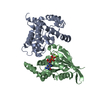
Mass: 24.305 Da / Num. of mol.: 1 / Source method: obtained synthetically / Formula: Mg
#4: Chemical ChemComp-GDP / GUANOSINE-5'-DIPHOSPHATE


Type: RNA linking / Mass: 443.201 Da / Num. of mol.: 1 / Source method: obtained synthetically / Formula: C10H15N5O11P2 / Comment: GDP, energy-carrying molecule*YM
#5: Chemical ChemComp-AF3 / ALUMINUM FLUORIDE


Mass: 83.977 Da / Num. of mol.: 1 / Source method: obtained synthetically / Formula: AlF3
#6: Water ChemComp-HOH / water


Mass: 18.015 Da / Num. of mol.: 103 / Source method: isolated from a natural source / Formula: H2O

-
Details

Has protein modificationY
Sequence detailsThis sequence is based on GenBank BAA90247.

-
Experimental details

-
Experiment

ExperimentMethod: X-RAY DIFFRACTION

-
Sample preparation

CrystalDensity Matthews: 1.97 Å3/Da / Density % sol: 37.48 %
Crystal growTemperature: 293 K / Method: vapor diffusion, sitting drop / pH: 5.5 / Details: PEG3350, Bis-Tris

-
Data collection

DiffractionMean temperature: 100 K
Diffraction sourceSource: SYNCHROTRON / Site: Photon Factory / Beamline: BL-5A / Wavelength: 1 Å
DetectorType: ADSC QUANTUM 315r / Detector: CCD / Date: Jan 28, 2009
RadiationProtocol: SINGLE WAVELENGTH / Monochromatic (M) / Laue (L): M / Scattering type: x-ray
Radiation wavelengthWavelength: 1 Å / Relative weight: 1
ReflectionResolution: 2.5→50 Å / Num. obs: 12204 / % possible obs: 99.9 % / Redundancy: 3.7 % / Biso Wilson estimate: 26 Å2 / Net I/σ(I): 13.2
Reflection shellResolution: 2.5→2.59 Å / Redundancy: 3.7 % / Mean I/σ(I) obs: 3.3 / Rsym value: 0.439 / % possible all: 100

-
Processing

Software
NameVersionClassification
CNS1.1refinement
HKL-2000data reduction
HKL-2000data scaling
MOLREPphasing
RefinementMethod to determine structure: MOLECULAR REPLACEMENT
Starting model: 2OVJ, 1GRN
Resolution: 2.5→32.39 Å / Rfactor Rfree error: 0.011 / Data cutoff high absF: 919676.15 / Data cutoff low absF: 0 / Isotropic thermal model: RESTRAINED / Cross valid method: THROUGHOUT / σ(F): 0
RfactorNum. reflection% reflectionSelection details
Rfree0.263 623 5.1 %RANDOM
Rwork0.198 ---
obs0.198 12153 99.9 %-
Solvent computationSolvent model: FLAT MODEL / Bsol: 26.1256 Å2 / ksol: 0.3201 e/Å3
Displacement parametersBiso mean: 36.6 Å2
Baniso -1Baniso -2Baniso -3
1-21.32 Å20 Å2-9.22 Å2
2---7.92 Å20 Å2
3----13.4 Å2
Refine analyze
FreeObs
Luzzati coordinate error0.41 Å0.29 Å
Luzzati d res low-5 Å
Luzzati sigma a0.5 Å0.38 Å
Refinement stepCycle: 1 / Resolution: 2.5→32.39 Å
ProteinNucleic acidLigandSolventTotal
Num. atoms3080 1613 32 103 4828
Refine LS restraints
Refine-IDTypeDev idealDev ideal target
X-RAY DIFFRACTIONc_bond_d0.007
X-RAY DIFFRACTIONc_bond_d_na
X-RAY DIFFRACTIONc_bond_d_prot
X-RAY DIFFRACTIONc_angle_d
X-RAY DIFFRACTIONc_angle_d_na
X-RAY DIFFRACTIONc_angle_d_prot
X-RAY DIFFRACTIONc_angle_deg1.3
X-RAY DIFFRACTIONc_angle_deg_na
X-RAY DIFFRACTIONc_angle_deg_prot
X-RAY DIFFRACTIONc_dihedral_angle_d21.5
X-RAY DIFFRACTIONc_dihedral_angle_d_na
X-RAY DIFFRACTIONc_dihedral_angle_d_prot
X-RAY DIFFRACTIONc_improper_angle_d1.01
X-RAY DIFFRACTIONc_improper_angle_d_na
X-RAY DIFFRACTIONc_improper_angle_d_prot
X-RAY DIFFRACTIONc_mcbond_it1.361.5
X-RAY DIFFRACTIONc_mcangle_it2.312
X-RAY DIFFRACTIONc_scbond_it2.072
X-RAY DIFFRACTIONc_scangle_it3.182.5
LS refinement shellResolution: 2.5→2.66 Å / Rfactor Rfree error: 0.038 / Total num. of bins used: 6
RfactorNum. reflection% reflection
Rfree0.378 98 4.9 %
Rwork0.291 1913 -
obs--99.9 %
Xplor file
Refine-IDSerial noParam fileTopol file
X-RAY DIFFRACTION1CNS_TOPPAR/protein_rep.paramCNS_TOPPAR/protein.top
X-RAY DIFFRACTION2CNS_TOPPAR/water_rep.paramCNS_TOPPAR/water.top
X-RAY DIFFRACTION3CNS_TOPPAR/ion.paramCNS_TOPPAR/ion.top
X-RAY DIFFRACTION4af3.paramaf3.top
X-RAY DIFFRACTION5GDP.paramGDP.top

+
About Yorodumi

-
News

-
Feb 9, 2022. New format data for meta-information of EMDB entries

New format data for meta-information of EMDB entries

  • Version 3 of the EMDB header file is now the official format.
  • The previous official version 1.9 will be removed from the archive.

Related info.:EMDB header

External links:wwPDB to switch to version 3 of the EMDB data model

-
Aug 12, 2020. Covid-19 info

Covid-19 info

URL: https://pdbj.org/emnavi/covid19.php

New page: Covid-19 featured information page in EM Navigator.

Related info.:Covid-19 info / Mar 5, 2020. Novel coronavirus structure data

+
Mar 5, 2020. Novel coronavirus structure data

Novel coronavirus structure data

Related info.:Yorodumi Speices / Aug 12, 2020. Covid-19 info

External links:COVID-19 featured content - PDBj / Molecule of the Month (242):Coronavirus Proteases

+
Jan 31, 2019. EMDB accession codes are about to change! (news from PDBe EMDB page)

EMDB accession codes are about to change! (news from PDBe EMDB page)

  • The allocation of 4 digits for EMDB accession codes will soon come to an end. Whilst these codes will remain in use, new EMDB accession codes will include an additional digit and will expand incrementally as the available range of codes is exhausted. The current 4-digit format prefixed with “EMD-” (i.e. EMD-XXXX) will advance to a 5-digit format (i.e. EMD-XXXXX), and so on. It is currently estimated that the 4-digit codes will be depleted around Spring 2019, at which point the 5-digit format will come into force.
  • The EM Navigator/Yorodumi systems omit the EMD- prefix.

Related info.:Q: What is EMD? / ID/Accession-code notation in Yorodumi/EM Navigator

External links:EMDB Accession Codes are Changing Soon! / Contact to PDBj

+
Jul 12, 2017. Major update of PDB

Major update of PDB

  • wwPDB released updated PDB data conforming to the new PDBx/mmCIF dictionary.
  • This is a major update changing the version number from 4 to 5, and with Remediation, in which all the entries are updated.
  • In this update, many items about electron microscopy experimental information are reorganized (e.g. em_software).
  • Now, EM Navigator and Yorodumi are based on the updated data.

External links:wwPDB Remediation / Enriched Model Files Conforming to OneDep Data Standards Now Available in the PDB FTP Archive

-
Yorodumi

Thousand views of thousand structures

  • Yorodumi is a browser for structure data from EMDB, PDB, SASBDB, etc.
  • This page is also the successor to EM Navigator detail page, and also detail information page/front-end page for Omokage search.
  • The word "yorodu" (or yorozu) is an old Japanese word meaning "ten thousand". "mi" (miru) is to see.

Related info.:EMDB / PDB / SASBDB / Comparison of 3 databanks / Yorodumi Search / Aug 31, 2016. New EM Navigator & Yorodumi / Yorodumi Papers / Jmol/JSmol / Function and homology information / Changes in new EM Navigator and Yorodumi

Read more