 Movie
Movie Controller

[English] 日本語
 Yorodumi
- PDB-2etw: Principles of protein-DNA recognition revealed in the structural ... -
+ Open data

- Basic information
| Entry | Database: PDB / ID: 2etw | ||||||
|---|---|---|---|---|---|---|---|
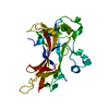
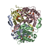
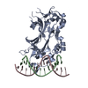
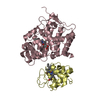
| Title | Principles of protein-DNA recognition revealed in the structural analysis of Ndt80-MSE DNA complexes | ||||||
 Components Components |
| ||||||
 Keywords Keywords | CELL CYCLE/DNA / beta sandwich / Ig-fold / B-DNA / CELL CYCLE-DNA COMPLEX | ||||||
| Function / homology |  Function and homology information Function and homology informationsporulation / nuclear chromosome / meiotic cell cycle / sequence-specific DNA binding / DNA-binding transcription factor activity / cell division / positive regulation of transcription by RNA polymerase II Similarity search - Function | ||||||
| Biological species |  | ||||||
| Method |  X-RAY DIFFRACTION / SYNCHROTRON / FOURIER SYNTHESIS / Resolution: 1.67 Å X-RAY DIFFRACTION / SYNCHROTRON / FOURIER SYNTHESIS / Resolution: 1.67 Å | ||||||
 Authors Authors | Lamoureux, J.S. / Glover, J.N. | ||||||
 Citation Citation | Journal: Structure / Year: 2006 Title: Principles of Protein-DNA Recognition Revealed in the Structural Analysis of Ndt80-MSE DNA Complexes. Authors: Lamoureux, J.S. / Glover, J.N. | ||||||
| History |
|
- Structure visualization
| Structure viewer | Molecule: MolmilJmol/JSmol |
|---|
- Downloads & links
-Download
| PDBx/mmCIF format | 2etw.cif.gz | 102.7 KB | Display | PDBx/mmCIF format |
|---|---|---|---|---|
| PDB format | pdb2etw.ent.gz | 73.7 KB | Display | PDB format |
| PDBx/mmJSON format | 2etw.json.gz | Tree view | PDBx/mmJSON format | |
| Others |  Other downloads Other downloads |
-Validation report
| Arichive directory | https://data.pdbj.org/pub/pdb/validation_reports/et/2etwftp://data.pdbj.org/pub/pdb/validation_reports/et/2etw | HTTPS FTP |
|---|
-Related structure data
| Related structure data | 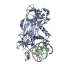 2euvC 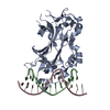 2euwC 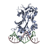 2euxC 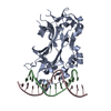 2euzC  2evfC  2evgC  2evhC  2eviC  2evjC  1mnnS S: Starting model for refinement C: citing same article ( |
|---|---|
| Similar structure data |


-Links
 PDBj
PDBj


- Assembly
| Deposited unit | 
| ||||||||
|---|---|---|---|---|---|---|---|---|---|
| 1 |
| ||||||||
| Unit cell |
|
-Components
| #1: DNA chain | Mass: 4226.797 Da / Num. of mol.: 1 / Source method: obtained synthetically / Details: vG1C MSE DNA strand 1 |
|---|---|
| #2: DNA chain | Mass: 4332.809 Da / Num. of mol.: 1 / Source method: obtained synthetically / Details: vG1C MSE DNA strand 2 |
| #3: Protein | Mass: 39590.527 Da / Num. of mol.: 1 / Fragment: Ndt80 DNA-binding Domain Source method: isolated from a genetically manipulated source Source: (gene. exp.) Gene: NDT80 / Plasmid: PGEX-6P1 / Species (production host): Escherichia coli / Production host:  |
| #4: Water | ChemComp-HOH /  Mass: 18.015 Da / Num. of mol.: 331 / Source method: isolated from a natural source / Formula: H2O Mass: 18.015 Da / Num. of mol.: 331 / Source method: isolated from a natural source / Formula: H2O |
-Experimental details
-Experiment
| Experiment | Method: X-RAY DIFFRACTION / Number of used crystals: 1 |
|---|
- Sample preparation
| Crystal | Density Matthews: 2.31 Å3/Da / Density % sol: 46.79 % | ||||||||||||||||||||||||||||||||||||||||
|---|---|---|---|---|---|---|---|---|---|---|---|---|---|---|---|---|---|---|---|---|---|---|---|---|---|---|---|---|---|---|---|---|---|---|---|---|---|---|---|---|---|
| Crystal grow | Temperature: 295 K / Method: vapor diffusion, hanging drop / pH: 7 Details: 30% PEG 400, 50 mM bis-tris-propane pH 7.0, 100 mM NaCl, 50 mM CaCl2, and 1 mM DTT. 10mg/ml protein 1:1 molar ration with duplex DNA , VAPOR DIFFUSION, HANGING DROP, temperature 295K | ||||||||||||||||||||||||||||||||||||||||
| Components of the solutions |
|
-Data collection
| Diffraction | Mean temperature: 105 K |
|---|---|
| Diffraction source | Source: SYNCHROTRON / Site: ALS  / Beamline: 8.3.1 / Wavelength: 1.072158 Å / Beamline: 8.3.1 / Wavelength: 1.072158 Å |
| Detector | Type: ADSC QUANTUM 210 / Detector: CCD / Date: Jul 14, 2003 |
| Radiation | Monochromator: DOUBLE CRYSTAL / Protocol: SINGLE WAVELENGTH / Monochromatic (M) / Laue (L): M / Scattering type: x-ray |
| Radiation wavelength | Wavelength: 1.072158 Å / Relative weight: 1 |
| Reflection | Resolution: 1.67→23.57 Å / Num. all: 51821 / Num. obs: 51821 / % possible obs: 99 % / Observed criterion σ(F): 0 / Observed criterion σ(I): 0 / Redundancy: 4.7 % / Rmerge(I) obs: 0.043 / Rsym value: 0.043 / Net I/σ(I): 10.5 |
| Reflection shell | Resolution: 1.67→1.76 Å / % possible obs: 98.3 % / Redundancy: 2.7 % / Rmerge(I) obs: 0.176 / Mean I/σ(I) obs: 3.8 / Num. measured obs: 7464 / Rsym value: 0.176 / % possible all: 98.3 |
- Processing
| Software |
| ||||||||||||||||||||||||||||||||||||||||||||||||||||||||||||||||||||||||||||||||||||||||||||||||||||
|---|---|---|---|---|---|---|---|---|---|---|---|---|---|---|---|---|---|---|---|---|---|---|---|---|---|---|---|---|---|---|---|---|---|---|---|---|---|---|---|---|---|---|---|---|---|---|---|---|---|---|---|---|---|---|---|---|---|---|---|---|---|---|---|---|---|---|---|---|---|---|---|---|---|---|---|---|---|---|---|---|---|---|---|---|---|---|---|---|---|---|---|---|---|---|---|---|---|---|---|---|---|
| Refinement | Method to determine structure: FOURIER SYNTHESIS Starting model: 1MNN.pdb without water molecules and DNA at position -1,1,2 of the MSE. Resolution: 1.67→23.57 Å / Cor.coef. Fo:Fc: 0.962 / Cor.coef. Fo:Fc free: 0.958 / SU B: 1.594 / SU ML: 0.055 / Cross valid method: THROUGHOUT / σ(F): 0 / ESU R: 0.095 / ESU R Free: 0.089 / Stereochemistry target values: MAXIMUM LIKELIHOOD / Details: HYDROGENS HAVE BEEN ADDED IN THE RIDING POSITIONS
| ||||||||||||||||||||||||||||||||||||||||||||||||||||||||||||||||||||||||||||||||||||||||||||||||||||
| Solvent computation | Ion probe radii: 0.8 Å / Shrinkage radii: 0.8 Å / VDW probe radii: 1.4 Å / Solvent model: BABINET MODEL WITH MASK | ||||||||||||||||||||||||||||||||||||||||||||||||||||||||||||||||||||||||||||||||||||||||||||||||||||
| Displacement parameters | Biso mean: 17.272 Å2
| ||||||||||||||||||||||||||||||||||||||||||||||||||||||||||||||||||||||||||||||||||||||||||||||||||||
| Refinement step | Cycle: LAST / Resolution: 1.67→23.57 Å
| ||||||||||||||||||||||||||||||||||||||||||||||||||||||||||||||||||||||||||||||||||||||||||||||||||||
| Refine LS restraints |
| ||||||||||||||||||||||||||||||||||||||||||||||||||||||||||||||||||||||||||||||||||||||||||||||||||||
| LS refinement shell | Resolution: 1.67→1.713 Å / Total num. of bins used: 20
|
