- PDB-3bwv: Crystal structure of deoxyribonucleotidase-like protein (NP_76406... -
+
Open data
ID or keywords:
Loading...
-
Basic information
Entry
Database: PDB / ID: 3bwv
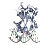
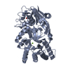
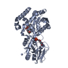
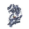
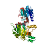
Title
Crystal structure of deoxyribonucleotidase-like protein (NP_764060.1) from Staphylococcus epidermidis ATCC 12228 at 1.55 A resolution
Components
Putative 5'(3')-deoxyribonucleotidase
Keywords
HYDROLASE / NP_764060.1 / deoxyribonucleotidase-like protein / Structural Genomics / Joint Center for Structural Genomics / JCSG / Protein Structure Initiative / PSI-2
Function / homology
Function and homology information
pyrimidine deoxyribonucleotide catabolic process / Hydrolases; Acting on ester bonds; Phosphoric-monoester hydrolases / 5'-nucleotidase activity / metal ion binding Similarity search - Function
5'(3')-deoxyribonucleotidase / 5' nucleotidase, deoxy (Pyrimidine), cytosolic type C protein (NT5C) / Deoxyribonucleotidase; domain 2 / Ribonucleotide Reductase Protein R1; domain 1 / HAD superfamily/HAD-like / HAD superfamily / HAD-like superfamily / Rossmann fold / Orthogonal Bundle / 3-Layer(aba) Sandwich ...5'(3')-deoxyribonucleotidase / 5' nucleotidase, deoxy (Pyrimidine), cytosolic type C protein (NT5C) / Deoxyribonucleotidase; domain 2 / Ribonucleotide Reductase Protein R1; domain 1 / HAD superfamily/HAD-like / HAD superfamily / HAD-like superfamily / Rossmann fold / Orthogonal Bundle / 3-Layer(aba) Sandwich / Mainly Alpha / Alpha Beta Similarity search - Domain/homology
SEQUENCE THE CONSTRUCT USED FOR REFINEMENT WAS EXPRESSED WITH A PURIFICATION TAG ... SEQUENCE THE CONSTRUCT USED FOR REFINEMENT WAS EXPRESSED WITH A PURIFICATION TAG MGSDKIHHHHHHENLYFQG. THE TAG WAS REMOVED WITH TEV PROTEASE LEAVING ONLY A GLYCINE (0) FOLLOWED BY THE TARGET SEQUENCE.
Type: MARMOSAIC 325 mm CCD / Detector: CCD / Date: Dec 9, 2007 / Details: Flat mirror (vertical focusing)
Radiation
Monochromator: Single crystal Si(111) bent (horizontal focusing) Protocol: MAD / Monochromatic (M) / Laue (L): M / Scattering type: x-ray
Radiation wavelength
ID
Wavelength (Å)
Relative weight
1
0.91837
1
2
0.97908
1
3
0.97959
1
Reflection
Resolution: 1.54→29.235 Å / Num. obs: 43195 / % possible obs: 92.8 % / Observed criterion σ(I): -3 / Biso Wilson estimate: 27.438 Å2 / Rmerge(I) obs: 0.067 / Net I/σ(I): 4.92
Reflection shell
Resolution (Å)
Rmerge(I) obs
Mean I/σ(I) obs
Num. measured obs
Num. unique obs
Diffraction-ID
% possible all
1.54-1.6
0.529
1.2
6168
7578
1
75.2
1.6-1.66
0.434
1.5
7022
8058
1
94
1.66-1.73
0.351
1.8
7184
8134
1
94.3
1.73-1.83
0.25
2.4
8502
9608
1
94.8
1.83-1.94
0.168
3.4
7322
8336
1
94.9
1.94-2.09
0.111
4.8
7646
8767
1
95
2.09-2.3
0.082
6.3
7638
8880
1
95.6
2.3-2.63
0.07
7.4
7322
8785
1
95.9
2.63-3.31
0.059
9
6960
8857
1
95.5
3.31-29.235
0.045
10.4
6650
8811
1
94.7
-
Phasing
Phasing
Method: MAD
-
Processing
Software
Name
Version
Classification
NB
REFMAC
5.4.0066
refinement
PHENIX
refinement
SOLVE
phasing
MolProbity
3beta29
modelbuilding
XSCALE
datascaling
PDB_EXTRACT
3
dataextraction
MAR345
CCD
datacollection
XDS
datareduction
Refinement
Method to determine structure: MAD / Resolution: 1.55→29.235 Å / Cor.coef. Fo:Fc: 0.968 / Cor.coef. Fo:Fc free: 0.946 / SU B: 4.605 / SU ML: 0.079 / TLS residual ADP flag: LIKELY RESIDUAL / Cross valid method: THROUGHOUT / σ(F): 0 / ESU R: 0.092 / ESU R Free: 0.095 Stereochemistry target values: MAXIMUM LIKELIHOOD WITH PHASES Details: 1. HYDROGENS HAVE BEEN ADDED IN THE RIDING POSITIONS. 2. A MET-INHIBITION PROTOCOL WAS USED FOR SELENOMETHIONINE INCORPORATION DURING PROTEIN EXPRESSION. THE OCCUPANCY OF THE SE ATOMS IN THE ...Details: 1. HYDROGENS HAVE BEEN ADDED IN THE RIDING POSITIONS. 2. A MET-INHIBITION PROTOCOL WAS USED FOR SELENOMETHIONINE INCORPORATION DURING PROTEIN EXPRESSION. THE OCCUPANCY OF THE SE ATOMS IN THE MSE RESIDUES WAS REDUCED TO 0.75 FOR THE REDUCED SCATTERING POWER DUE TO PARTIAL S-MET INCORPORATION. 3. ATOM RECORD CONTAINS RESIDUAL B FACTORS ONLY. 4. MG AND CL MODELED BASED ON GEOMETRY AND CRYSTALLIZATION CONDITIONS.
Rfactor
Num. reflection
% reflection
Selection details
Rfree
0.223
2176
5 %
RANDOM
Rwork
0.182
-
-
-
obs
0.184
43194
94.9 %
-
Solvent computation
Ion probe radii: 0.8 Å / Shrinkage radii: 0.8 Å / VDW probe radii: 1.2 Å / Solvent model: MASK
Displacement parameters
Biso mean: 19.268 Å2
Baniso -1
Baniso -2
Baniso -3
1-
0.12 Å2
-0.21 Å2
-0.58 Å2
2-
-
-0.79 Å2
0.77 Å2
3-
-
-
0.54 Å2
Refinement step
Cycle: LAST / Resolution: 1.55→29.235 Å
Protein
Nucleic acid
Ligand
Solvent
Total
Num. atoms
2700
0
4
181
2885
Refine LS restraints
Refine-ID
Type
Dev ideal
Dev ideal target
Number
X-RAY DIFFRACTION
r_bond_refined_d
0.017
0.022
2819
X-RAY DIFFRACTION
r_bond_other_d
0.003
0.02
1879
X-RAY DIFFRACTION
r_angle_refined_deg
1.634
1.941
3832
X-RAY DIFFRACTION
r_angle_other_deg
1.044
3
4576
X-RAY DIFFRACTION
r_dihedral_angle_1_deg
5.971
5
349
X-RAY DIFFRACTION
r_dihedral_angle_2_deg
35.647
24.167
144
X-RAY DIFFRACTION
r_dihedral_angle_3_deg
13.772
15
476
X-RAY DIFFRACTION
r_dihedral_angle_4_deg
21.593
15
16
X-RAY DIFFRACTION
r_chiral_restr
0.104
0.2
422
X-RAY DIFFRACTION
r_gen_planes_refined
0.007
0.02
3154
X-RAY DIFFRACTION
r_gen_planes_other
0.001
0.02
602
X-RAY DIFFRACTION
r_mcbond_it
1.004
1.5
1698
X-RAY DIFFRACTION
r_mcbond_other
0.301
1.5
680
X-RAY DIFFRACTION
r_mcangle_it
1.748
2
2748
X-RAY DIFFRACTION
r_scbond_it
3.105
3
1121
X-RAY DIFFRACTION
r_scangle_it
4.812
4.5
1074
LS refinement shell
Resolution: 1.55→1.59 Å / Total num. of bins used: 20
Rfactor
Num. reflection
% reflection
Rfree
0.309
124
-
Rwork
0.294
2792
-
all
-
2916
-
obs
-
-
86.76 %
Refinement TLS params.
Method: refined / Refine-ID: X-RAY DIFFRACTION
ID
L11 (°2)
L12 (°2)
L13 (°2)
L22 (°2)
L23 (°2)
L33 (°2)
S11 (Å °)
S12 (Å °)
S13 (Å °)
S21 (Å °)
S22 (Å °)
S23 (Å °)
S31 (Å °)
S32 (Å °)
S33 (Å °)
T11 (Å2)
T12 (Å2)
T13 (Å2)
T22 (Å2)
T23 (Å2)
T33 (Å2)
Origin x (Å)
Origin y (Å)
Origin z (Å)
1
2.6255
-0.0568
-0.2049
1.8546
-0.5095
1.5402
0.0451
-0.0373
-0.201
0.0031
0.0445
0.1524
0.0587
-0.0611
-0.0896
-0.0429
-0.0036
-0.0008
-0.0424
-0.0058
-0.0632
16.4425
19.0579
44.5968
2
0
0
0
0
0
0
0
0
0
0
0
0
0
0
0
-0.0237
-0.0243
-0.0392
-0.0504
-0.0117
0.0036
15.9106
15.285
41.5251
3
2.1807
-0.4027
0.1575
1.1639
-0.5759
1.3943
0.0158
-0.0169
0.0008
-0.0287
0.0326
0.1476
-0.0099
-0.0384
-0.0483
-0.0012
-0.0037
0.0035
-0.0453
-0.0102
-0.0388
16.809
35.1872
19.5333
4
0
0
0
0
0
0
0
0
0
0
0
0
0
0
0
0.029
0.007
0.0345
-0.0212
-0.0936
-0.0711
16.3695
38.4765
22.5175
Refinement TLS group
ID
Refine-ID
Refine TLS-ID
Auth asym-ID
Label asym-ID
Auth seq-ID
Label seq-ID
1
X-RAY DIFFRACTION
1
A
A
2 - 178
3 - 179
2
X-RAY DIFFRACTION
2
A
C
300
1
3
X-RAY DIFFRACTION
3
B
B
3 - 178
4 - 179
4
X-RAY DIFFRACTION
4
B
D
300
1
+
About Yorodumi
-
News
-
Feb 9, 2022. New format data for meta-information of EMDB entries
New format data for meta-information of EMDB entries
Version 3 of the EMDB header file is now the official format.
The previous official version 1.9 will be removed from the archive.
In the structure databanks used in Yorodumi, some data are registered as the other names, "COVID-19 virus" and "2019-nCoV". Here are the details of the virus and the list of structure data.
Jan 31, 2019. EMDB accession codes are about to change! (news from PDBe EMDB page)
EMDB accession codes are about to change! (news from PDBe EMDB page)
The allocation of 4 digits for EMDB accession codes will soon come to an end. Whilst these codes will remain in use, new EMDB accession codes will include an additional digit and will expand incrementally as the available range of codes is exhausted. The current 4-digit format prefixed with “EMD-” (i.e. EMD-XXXX) will advance to a 5-digit format (i.e. EMD-XXXXX), and so on. It is currently estimated that the 4-digit codes will be depleted around Spring 2019, at which point the 5-digit format will come into force.
The EM Navigator/Yorodumi systems omit the EMD- prefix.
Related info.:Q: What is EMD? / ID/Accession-code notation in Yorodumi/EM Navigator
Yorodumi is a browser for structure data from EMDB, PDB, SASBDB, etc.
This page is also the successor to EM Navigator detail page, and also detail information page/front-end page for Omokage search.
The word "yorodu" (or yorozu) is an old Japanese word meaning "ten thousand". "mi" (miru) is to see.
Related info.:EMDB / PDB / SASBDB / Comparison of 3 databanks / Yorodumi Search / Aug 31, 2016. New EM Navigator & Yorodumi / Yorodumi Papers / Jmol/JSmol / Function and homology information / Changes in new EM Navigator and Yorodumi
 Movie
Movie Controller

 Yorodumi
 Open data
 Basic information
 Components
 Keywords
 Function and homology information
 Staphylococcus epidermidis (bacteria)
 X-RAY DIFFRACTION /
  Authors
 Citation
 Structure visualization
 Downloads & links
 Other downloads
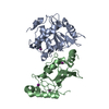


 PDBj
PDBj Assembly
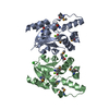

 Mass: 24.305 Da / Num. of mol.: 2 / Source method: obtained synthetically / Formula: Mg
 Mass: 35.453 Da / Num. of mol.: 2 / Source method: obtained synthetically / Formula: Cl
 Mass: 18.015 Da / Num. of mol.: 181 / Source method: isolated from a natural source / Formula: H2O
 Sample preparation
 / Beamline: BL11-1 / Wavelength: 0.91837, 0.97908, 0.97959
 Processing