 Movie
Movie Controller

[English] 日本語
 Yorodumi
- PDB-3w31: Structual basis for the recognition of Ubc13 by the Shigella flex... -
+ Open data

- Basic information
| Entry | Database: PDB / ID: 3w31 | ||||||
|---|---|---|---|---|---|---|---|
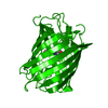
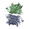
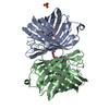
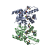
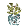
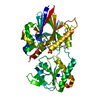
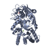
| Title | Structual basis for the recognition of Ubc13 by the Shigella flexneri effector OspI | ||||||
 Components Components |
| ||||||
 Keywords Keywords | IMMUNE SYSTEM / Type 3 secretion system / Effector / Deamidation | ||||||
| Function / homology |  Function and homology information Function and homology informationUBC13-MMS2 complex / ubiquitin conjugating enzyme complex / ubiquitin-protein transferase activator activity / positive regulation of protein K63-linked ubiquitination / DNA double-strand break processing / DNA damage tolerance / E2 ubiquitin-conjugating enzyme / symbiont-mediated suppression of host TRAF-mediated signal transduction / positive regulation of double-strand break repair / ubiquitin conjugating enzyme activity ...UBC13-MMS2 complex / ubiquitin conjugating enzyme complex / ubiquitin-protein transferase activator activity / positive regulation of protein K63-linked ubiquitination / DNA double-strand break processing / DNA damage tolerance / E2 ubiquitin-conjugating enzyme / symbiont-mediated suppression of host TRAF-mediated signal transduction / positive regulation of double-strand break repair / ubiquitin conjugating enzyme activity / positive regulation of intracellular signal transduction / protein monoubiquitination / protein K63-linked ubiquitination / ubiquitin ligase complex / regulation of DNA repair / negative regulation of TORC1 signaling / : / IRAK1 recruits IKK complex / IRAK1 recruits IKK complex upon TLR7/8 or 9 stimulation / TRAF6 mediated IRF7 activation in TLR7/8 or 9 signaling / antiviral innate immune response / positive regulation of DNA repair / TICAM1, RIP1-mediated IKK complex recruitment / JNK (c-Jun kinases) phosphorylation and activation mediated by activated human TAK1 / IKK complex recruitment mediated by RIP1 / PINK1-PRKN Mediated Mitophagy / activated TAK1 mediates p38 MAPK activation / ubiquitin binding / Nonhomologous End-Joining (NHEJ) / NOD1/2 Signaling Pathway / TAK1-dependent IKK and NF-kappa-B activation / G2/M DNA damage checkpoint / double-strand break repair via homologous recombination / ISG15 antiviral mechanism / CLEC7A (Dectin-1) signaling / FCERI mediated NF-kB activation / Formation of Incision Complex in GG-NER / Interleukin-1 signaling / protein polyubiquitination / Aggrephagy / ubiquitin-protein transferase activity / T cell receptor signaling pathway / Downstream TCR signaling / Antigen processing: Ubiquitination & Proteasome degradation / E3 ubiquitin ligases ubiquitinate target proteins / Recruitment and ATM-mediated phosphorylation of repair and signaling proteins at DNA double strand breaks / Processing of DNA double-strand break ends / proteasome-mediated ubiquitin-dependent protein catabolic process / positive regulation of canonical NF-kappaB signal transduction / protein ubiquitination / ubiquitin protein ligase binding / SARS-CoV-2 activates/modulates innate and adaptive immune responses / protein-containing complex / RNA binding / extracellular exosome / nucleoplasm / ATP binding / nucleus / cytosol / cytoplasm Similarity search - Function | ||||||
| Biological species |  Shigella flexneri (bacteria) Shigella flexneri (bacteria) Homo sapiens (human) Homo sapiens (human) | ||||||
| Method |  X-RAY DIFFRACTION / SYNCHROTRON / MOLECULAR REPLACEMENT / Resolution: 2.96 Å X-RAY DIFFRACTION / SYNCHROTRON / MOLECULAR REPLACEMENT / Resolution: 2.96 Å | ||||||
 Authors Authors | Nishide, A. / Kim, M. / Takagi, K. / Sasakawa, C. / Mizushima, T. | ||||||
 Citation Citation | Journal: J.Mol.Biol. / Year: 2013 Title: Structural basis for the recognition of Ubc13 by the Shigella flexneri effector OspI. Authors: Nishide, A. / Kim, M. / Takagi, K. / Himeno, A. / Sanada, T. / Sasakawa, C. / Mizushima, T. | ||||||
| History |
|
- Structure visualization
| Structure viewer | Molecule: MolmilJmol/JSmol |
|---|
- Downloads & links
-Download
| PDBx/mmCIF format | 3w31.cif.gz | 80.5 KB | Display | PDBx/mmCIF format |
|---|---|---|---|---|
| PDB format | pdb3w31.ent.gz | 59.9 KB | Display | PDB format |
| PDBx/mmJSON format | 3w31.json.gz | Tree view | PDBx/mmJSON format | |
| Others |  Other downloads Other downloads |
-Validation report
| Arichive directory | https://data.pdbj.org/pub/pdb/validation_reports/w3/3w31ftp://data.pdbj.org/pub/pdb/validation_reports/w3/3w31 | HTTPS FTP |
|---|
-Related structure data
| Related structure data |  3w30C 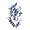 1jbbS 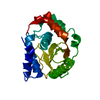 3b21S C: citing same article ( S: Starting model for refinement |
|---|---|
| Similar structure data |


-Links
 PDBj
PDBj


- Assembly
| Deposited unit | 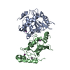
| ||||||||
|---|---|---|---|---|---|---|---|---|---|
| 1 |
| ||||||||
| Unit cell |
|
-Components
| #1: Protein | Mass: 24118.807 Da / Num. of mol.: 1 / Mutation: C62A Source method: isolated from a genetically manipulated source Source: (gene. exp.) Shigella flexneri (bacteria) / Gene: ORF169b, CP0209 / Production host: |
|---|---|
| #2: Protein | Mass: 17440.049 Da / Num. of mol.: 1 Source method: isolated from a genetically manipulated source Source: (gene. exp.) Homo sapiens (human) / Gene: UBE2N, BLU / Production host: |
| #3: Chemical | ChemComp-IOD /   Mass: 126.904 Da / Num. of mol.: 4 / Source method: obtained synthetically / Formula: I Mass: 126.904 Da / Num. of mol.: 4 / Source method: obtained synthetically / Formula: I |
-Experimental details
-Experiment
| Experiment | Method: X-RAY DIFFRACTION |
|---|
- Sample preparation
| Crystal | Density Matthews: 3.4 Å3/Da / Density % sol: 63.81 % |
|---|---|
| Crystal grow | Temperature: 293 K / Method: vapor diffusion, hanging drop / pH: 6.5 Details: 0.4M potassium iodide, 0.1M MES (pH6.5), 4mM DTT, 15% PEG 8000, VAPOR DIFFUSION, HANGING DROP, temperature 293K |
-Data collection
| Diffraction source | Source: SYNCHROTRON / Site: SPring-8  / Beamline: BL44XU / Wavelength: 0.9 Å / Beamline: BL44XU / Wavelength: 0.9 Å |
|---|---|
| Detector | Date: Dec 14, 2011 |
| Radiation | Protocol: SINGLE WAVELENGTH / Monochromatic (M) / Laue (L): M / Scattering type: x-ray |
| Radiation wavelength | Wavelength: 0.9 Å / Relative weight: 1 |
| Reflection | Resolution: 2.96→50 Å / Num. all: 12250 / Num. obs: 12193 / % possible obs: 99.8 % / Redundancy: 3.4 % / Rmerge(I) obs: 0.094 |
- Processing
| Software |
| ||||||||||||||||
|---|---|---|---|---|---|---|---|---|---|---|---|---|---|---|---|---|---|
| Refinement | Method to determine structure: MOLECULAR REPLACEMENT Starting model: 3B21, 1JBB Resolution: 2.96→33.1 Å
| ||||||||||||||||
| Displacement parameters |
| ||||||||||||||||
| Refinement step | Cycle: LAST / Resolution: 2.96→33.1 Å
|
