 Movie
Movie Controller

[English] 日本語
 Yorodumi
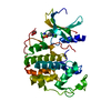
- PDB-1m6u: Crystal Structure of a Novel DNA-binding domain from Ndt80, a Tra... -
+ Open data

- Basic information
| Entry | Database: PDB / ID: 1m6u | ||||||
|---|---|---|---|---|---|---|---|
| Title | Crystal Structure of a Novel DNA-binding domain from Ndt80, a Transcriptional Activator Required for Meiosis in Yeast | ||||||
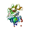
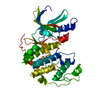
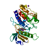
 Components Components | Ndt80 protein | ||||||
 Keywords Keywords | TRANSCRIPTION ACTIVATOR / yeast protein / DNA-binding / meiosis | ||||||
| Function / homology |  Function and homology information Function and homology informationsporulation / nuclear chromosome / meiotic cell cycle / sequence-specific DNA binding / DNA-binding transcription factor activity / cell division / positive regulation of transcription by RNA polymerase II Similarity search - Function | ||||||
| Biological species |  | ||||||
| Method |  X-RAY DIFFRACTION / MOLECULAR REPLACEMENT / Resolution: 2.3 Å X-RAY DIFFRACTION / MOLECULAR REPLACEMENT / Resolution: 2.3 Å | ||||||
 Authors Authors | Montano, S.P. / Cote, M.L. / Fingerman, I. / Pierce, M. / Vershon, A.K. / Georgiadis, M.M. | ||||||
 Citation Citation | Journal: Proc.Natl.Acad.Sci.USA / Year: 2002 Title: Crystal structure of the DNA-binding domain from Ndt80, a transcriptional activator required for meiosis in yeast Authors: Montano, S.P. / Cote, M.L. / Fingerman, I. / Pierce, M. / Vershon, A.K. / Georgiadis, M.M. | ||||||
| History |
| ||||||
| Remark 999 | SEQUENCE THE DISCREPANCIES ARE PRESENT DUE TO ERRORS IN PCR. |
- Structure visualization
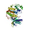
| Structure viewer | Molecule: MolmilJmol/JSmol |
|---|
- Downloads & links
-Download
| PDBx/mmCIF format | 1m6u.cif.gz | 116 KB | Display | PDBx/mmCIF format |
|---|---|---|---|---|
| PDB format | pdb1m6u.ent.gz | 89.5 KB | Display | PDB format |
| PDBx/mmJSON format | 1m6u.json.gz | Tree view | PDBx/mmJSON format | |
| Others |  Other downloads Other downloads |
-Validation report
| Arichive directory | https://data.pdbj.org/pub/pdb/validation_reports/m6/1m6uftp://data.pdbj.org/pub/pdb/validation_reports/m6/1m6u | HTTPS FTP |
|---|
-Related structure data
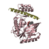
| Related structure data | 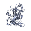 1m7uSC S: Starting model for refinement C: citing same article ( |
|---|---|
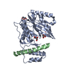
| Similar structure data |
-Links
 PDBj
PDBj


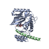
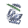
- Assembly
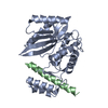
| Deposited unit | 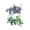
| ||||||||
|---|---|---|---|---|---|---|---|---|---|
| 1 | 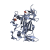
| ||||||||
| 2 | 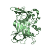
| ||||||||
| 3 | 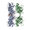
| ||||||||
| 4 | 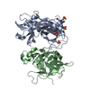
| ||||||||
| 5 | 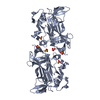
| ||||||||
| Unit cell |
|
-Components
| #1: Protein | Mass: 31307.455 Da / Num. of mol.: 2 / Fragment: DNA binding domain, residues 59-330 Source method: isolated from a genetically manipulated source Source: (gene. exp.) Gene: Ndt80 / Plasmid: pET15b / Production host:  #2: Chemical | ChemComp-SO4 /   Mass: 96.063 Da / Num. of mol.: 4 / Source method: obtained synthetically / Formula: SO4 Mass: 96.063 Da / Num. of mol.: 4 / Source method: obtained synthetically / Formula: SO4#3: Water | ChemComp-HOH / |  Mass: 18.015 Da / Num. of mol.: 110 / Source method: isolated from a natural source / Formula: H2O Mass: 18.015 Da / Num. of mol.: 110 / Source method: isolated from a natural source / Formula: H2O |
|---|
-Experimental details
-Experiment
| Experiment | Method: X-RAY DIFFRACTION / Number of used crystals: 1 |
|---|
- Sample preparation
| Crystal | Density Matthews: 2.31 Å3/Da / Density % sol: 46.78 % | ||||||||||||||||||||||||
|---|---|---|---|---|---|---|---|---|---|---|---|---|---|---|---|---|---|---|---|---|---|---|---|---|---|
| Crystal grow | Temperature: 298 K / Method: vapor diffusion, hanging drop / pH: 5.6 Details: lithium sulfate, citric acid, pH 5.6, VAPOR DIFFUSION, HANGING DROP, temperature 298K | ||||||||||||||||||||||||
| Crystal grow | *PLUS Details: Montano, S.P., (2002) Acta Crystallogr., D58, 2127. | ||||||||||||||||||||||||
| Components of the solutions | *PLUS
|
-Data collection
| Diffraction | Mean temperature: 108 K |
|---|---|
| Diffraction source | Source: ROTATING ANODE / Type: RIGAKU / Wavelength: 1.5418 Å |
| Detector | Type: RIGAKU RAXIS IV / Detector: IMAGE PLATE / Date: May 16, 2001 / Details: mirrors |
| Radiation | Monochromator: YALE MIRRORS / Protocol: SINGLE WAVELENGTH / Monochromatic (M) / Laue (L): M / Scattering type: x-ray |
| Radiation wavelength | Wavelength: 1.5418 Å / Relative weight: 1 |
| Reflection | Resolution: 2.3→50 Å / Num. all: 25573 / Num. obs: 25573 / % possible obs: 95.7 % / Observed criterion σ(F): 2 / Observed criterion σ(I): 2 / Redundancy: 5.6 % / Biso Wilson estimate: 43 Å2 / Rmerge(I) obs: 0.07 / Rsym value: 0.13 / Net I/σ(I): 15.8 |
| Reflection shell | Resolution: 2.3→2.38 Å / Redundancy: 3.2 % / Rmerge(I) obs: 0.24 / Mean I/σ(I) obs: 3.3 / Num. unique all: 2128 / % possible all: 84.9 |
| Reflection | *PLUS Rmerge(I) obs: 0.084 |
| Reflection shell | *PLUS % possible obs: 84.9 % / Rmerge(I) obs: 0.241 / Mean I/σ(I) obs: 3.4 |
- Processing
| Software |
| |||||||||||||||||||||||||
|---|---|---|---|---|---|---|---|---|---|---|---|---|---|---|---|---|---|---|---|---|---|---|---|---|---|---|
| Refinement | Method to determine structure: MOLECULAR REPLACEMENT Starting model: Ndt80, pdb entry 1M7U Resolution: 2.3→19.96 Å / Isotropic thermal model: Overall / Cross valid method: THROUGHOUT / σ(F): 0 / σ(I): 0 / Stereochemistry target values: Engh & Huber / Details: maximum likelihood
| |||||||||||||||||||||||||
| Refinement step | Cycle: LAST / Resolution: 2.3→19.96 Å
| |||||||||||||||||||||||||
| LS refinement shell | Resolution: 2.3→2.33 Å /
| |||||||||||||||||||||||||
| Refinement | *PLUS % reflection Rfree: 5 % / Rfactor Rfree: 0.271 / Rfactor Rwork: 0.226 | |||||||||||||||||||||||||
| Solvent computation | *PLUS | |||||||||||||||||||||||||
| Displacement parameters | *PLUS | |||||||||||||||||||||||||
| Refine LS restraints | *PLUS
| |||||||||||||||||||||||||
| LS refinement shell | *PLUS Rfactor Rfree: 0.43 / Rfactor Rwork: 0.33 |
