 Movie
Movie Controller

[English] 日本語
 Yorodumi
- PDB-6j4u: Structural basis of tubulin detyrosination by vasohibins-SVBP enz... -
+ Open data

- Basic information
| Entry | Database: PDB / ID: 6j4u | ||||||||||||
|---|---|---|---|---|---|---|---|---|---|---|---|---|---|
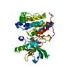
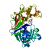
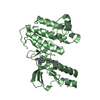
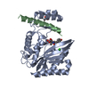
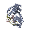
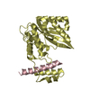
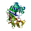
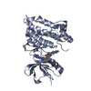
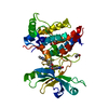
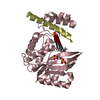
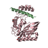
| Title | Structural basis of tubulin detyrosination by vasohibins-SVBP enzyme complex and functional implications | ||||||||||||
 Components Components |
| ||||||||||||
 Keywords Keywords | HYDROLASE / carboxypeptidase / tubulin / microtubule. | ||||||||||||
| Function / homology |  Function and homology information Function and homology informationregulation of metallopeptidase activity / tubulinyl-Tyr carboxypeptidase / tubulin-tyrosine carboxypeptidase activity / negative regulation of lymphangiogenesis / regulation of cellular senescence / Carboxyterminal post-translational modifications of tubulin / negative regulation of endothelial cell migration / labyrinthine layer blood vessel development / peptidase activator activity / negative regulation of endothelial cell proliferation ...regulation of metallopeptidase activity / tubulinyl-Tyr carboxypeptidase / tubulin-tyrosine carboxypeptidase activity / negative regulation of lymphangiogenesis / regulation of cellular senescence / Carboxyterminal post-translational modifications of tubulin / negative regulation of endothelial cell migration / labyrinthine layer blood vessel development / peptidase activator activity / negative regulation of endothelial cell proliferation / axon development / negative regulation of blood vessel endothelial cell migration / protein secretion / regulation of angiogenesis / metallocarboxypeptidase activity / negative regulation of protein ubiquitination / negative regulation of angiogenesis / response to wounding / apical part of cell / actin binding / angiogenesis / microtubule binding / cytoskeleton / regulation of cell cycle / endoplasmic reticulum / proteolysis / : / extracellular region / cytoplasm Similarity search - Function | ||||||||||||
| Biological species |  Homo sapiens (human) Homo sapiens (human) | ||||||||||||
| Method |  X-RAY DIFFRACTION / SYNCHROTRON / MOLECULAR REPLACEMENT / Resolution: 1.998 Å X-RAY DIFFRACTION / SYNCHROTRON / MOLECULAR REPLACEMENT / Resolution: 1.998 Å | ||||||||||||
 Authors Authors | Wang, N. / Bao, H. / Huang, H. | ||||||||||||
| Funding support |  China, 3items China, 3items
| ||||||||||||
 Citation Citation | Journal: Nat.Struct.Mol.Biol. / Year: 2019 Title: Structural basis of tubulin detyrosination by the vasohibin-SVBP enzyme complex. Authors: Wang, N. / Bosc, C. / Ryul Choi, S. / Boulan, B. / Peris, L. / Olieric, N. / Bao, H. / Krichen, F. / Chen, L. / Andrieux, A. / Olieric, V. / Moutin, M.J. / Steinmetz, M.O. / Huang, H. | ||||||||||||
| History |
|
- Structure visualization
| Structure viewer | Molecule: MolmilJmol/JSmol |
|---|
- Downloads & links
-Download
| PDBx/mmCIF format | 6j4u.cif.gz | 136.6 KB | Display | PDBx/mmCIF format |
|---|---|---|---|---|
| PDB format | pdb6j4u.ent.gz | 105.2 KB | Display | PDB format |
| PDBx/mmJSON format | 6j4u.json.gz | Tree view | PDBx/mmJSON format | |
| Others |  Other downloads Other downloads |
-Validation report
| Arichive directory | https://data.pdbj.org/pub/pdb/validation_reports/j4/6j4uftp://data.pdbj.org/pub/pdb/validation_reports/j4/6j4u | HTTPS FTP |
|---|
-Related structure data
| Related structure data |  6j4oSC  6j4pC  6j4qC  6j4sC 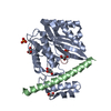 6j4vC  6qbyC S: Starting model for refinement C: citing same article ( |
|---|---|
| Similar structure data |


-Links
 PDBj
PDBj
- Assembly
| Deposited unit | 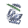
| ||||||||
|---|---|---|---|---|---|---|---|---|---|
| 1 |
| ||||||||
| Unit cell |
| ||||||||
| Components on special symmetry positions |
|
-Components
| #1: Protein | Mass: 29034.707 Da / Num. of mol.: 1 / Mutation: E71S/A72H/K79M Source method: isolated from a genetically manipulated source Source: (gene. exp.) Homo sapiens (human) / Gene: VASH1, KIAA1036, VASH / Plasmid: RSFdeut / Production host:  |
|---|---|
| #2: Protein | Mass: 7821.939 Da / Num. of mol.: 1 Source method: isolated from a genetically manipulated source Source: (gene. exp.) Homo sapiens (human) / Gene: SVBP, CCDC23 / Plasmid: RSFdeut / Production host: |
| #3: Water | ChemComp-HOH /  Mass: 18.015 Da / Num. of mol.: 238 / Source method: isolated from a natural source / Formula: H2O Mass: 18.015 Da / Num. of mol.: 238 / Source method: isolated from a natural source / Formula: H2O |
-Experimental details
-Experiment
| Experiment | Method: X-RAY DIFFRACTION / Number of used crystals: 1 |
|---|
- Sample preparation
| Crystal | Density Matthews: 2.76 Å3/Da / Density % sol: 55.48 % |
|---|---|
| Crystal grow | Temperature: 293 K / Method: evaporation / pH: 5 Details: by introducing three mutations E71S/A72H/K79M into V1c allowed us to solve the V1c-SVBP to high resolution. Well diffracting crystals of the mutant V1c-SVBP complex were obtained in 1.0 M ...Details: by introducing three mutations E71S/A72H/K79M into V1c allowed us to solve the V1c-SVBP to high resolution. Well diffracting crystals of the mutant V1c-SVBP complex were obtained in 1.0 M lithium chloride, 0.1 M citric acid, pH 5.0, 20% PEG 6000. PH range: 5 |
-Data collection
| Diffraction | Mean temperature: 100 K / Serial crystal experiment: N |
|---|---|
| Diffraction source | Source: SYNCHROTRON / Site: SSRF / Beamline: BL19U1 / Wavelength: 0.9789 Å |
| Detector | Type: DECTRIS PILATUS3 S 6M / Detector: PIXEL / Date: Jun 24, 2018 |
| Radiation | Protocol: SINGLE WAVELENGTH / Monochromatic (M) / Laue (L): M / Scattering type: x-ray |
| Radiation wavelength | Wavelength: 0.9789 Å / Relative weight: 1 |
| Reflection | Resolution: 1.998→50 Å / Num. obs: 28511 / % possible obs: 99.9 % / Redundancy: 12.4 % / Biso Wilson estimate: 39.4 Å2 / Rmerge(I) obs: 0.073 / Rpim(I) all: 0.023 / Net I/σ(I): 24.6 |
| Reflection shell | Resolution: 2→2.07 Å / Redundancy: 9.5 % / Rmerge(I) obs: 1 / Mean I/σ(I) obs: 1.2 / Num. unique obs: 2759 / CC1/2: 0.738 / Rpim(I) all: 0.358 / % possible all: 99.5 |
- Processing
| Software |
| ||||||||||||||||||||||||||||||||||||||||||||||||||||||||||||||||||||||||||||||||||||||||||||||||||||||||||||||||||||||||||||||||||||||||||||||||||||||
|---|---|---|---|---|---|---|---|---|---|---|---|---|---|---|---|---|---|---|---|---|---|---|---|---|---|---|---|---|---|---|---|---|---|---|---|---|---|---|---|---|---|---|---|---|---|---|---|---|---|---|---|---|---|---|---|---|---|---|---|---|---|---|---|---|---|---|---|---|---|---|---|---|---|---|---|---|---|---|---|---|---|---|---|---|---|---|---|---|---|---|---|---|---|---|---|---|---|---|---|---|---|---|---|---|---|---|---|---|---|---|---|---|---|---|---|---|---|---|---|---|---|---|---|---|---|---|---|---|---|---|---|---|---|---|---|---|---|---|---|---|---|---|---|---|---|---|---|---|---|---|---|
| Refinement | Method to determine structure: MOLECULAR REPLACEMENT Starting model: 6J4O Resolution: 1.998→47.695 Å / SU ML: 0.21 / Cross valid method: FREE R-VALUE / σ(F): 1.35 / Phase error: 21.23
| ||||||||||||||||||||||||||||||||||||||||||||||||||||||||||||||||||||||||||||||||||||||||||||||||||||||||||||||||||||||||||||||||||||||||||||||||||||||
| Solvent computation | Shrinkage radii: 0.9 Å / VDW probe radii: 1.11 Å | ||||||||||||||||||||||||||||||||||||||||||||||||||||||||||||||||||||||||||||||||||||||||||||||||||||||||||||||||||||||||||||||||||||||||||||||||||||||
| Refinement step | Cycle: LAST / Resolution: 1.998→47.695 Å
| ||||||||||||||||||||||||||||||||||||||||||||||||||||||||||||||||||||||||||||||||||||||||||||||||||||||||||||||||||||||||||||||||||||||||||||||||||||||
| Refine LS restraints |
| ||||||||||||||||||||||||||||||||||||||||||||||||||||||||||||||||||||||||||||||||||||||||||||||||||||||||||||||||||||||||||||||||||||||||||||||||||||||
| LS refinement shell |
| ||||||||||||||||||||||||||||||||||||||||||||||||||||||||||||||||||||||||||||||||||||||||||||||||||||||||||||||||||||||||||||||||||||||||||||||||||||||
| Refinement TLS params. | Method: refined / Refine-ID: X-RAY DIFFRACTION
| ||||||||||||||||||||||||||||||||||||||||||||||||||||||||||||||||||||||||||||||||||||||||||||||||||||||||||||||||||||||||||||||||||||||||||||||||||||||
| Refinement TLS group |
|
