Protocol: SINGLE WAVELENGTH / Monochromatic (M) / Laue (L): M / Scattering type: x-ray
Radiation wavelength
Wavelength: 0.966 Å / Relative weight: 1
Reflection
Resolution: 2.1→47.61 Å / Num. obs: 24696 / % possible obs: 99.7 % / Redundancy: 4.9 % / CC1/2: 0.99 / Rmerge(I) obs: 0.062 / Net I/σ(I): 12.8
Reflection shell
Resolution: 2.1→2.21 Å / Redundancy: 4.9 % / Rmerge(I) obs: 1.538 / Mean I/σ(I) obs: 1.1 / Num. unique obs: 3536 / CC1/2: 0.518 / % possible all: 51.8
-
Processing
Software
Name
Version
Classification
REFMAC
5.8.0238
refinement
PDB_EXTRACT
3.22
dataextraction
XDS
datareduction
SCALA
datascaling
CRANK2
phasing
Refinement
Method to determine structure: SAD / Resolution: 2.1→47.61 Å / Cor.coef. Fo:Fc: 0.965 / Cor.coef. Fo:Fc free: 0.96 / SU B: 11.353 / SU ML: 0.138 / Cross valid method: THROUGHOUT / ESU R: 0.171 / ESU R Free: 0.144 / Details: HYDROGENS HAVE BEEN ADDED IN THE RIDING POSITIONS
Rfactor
Num. reflection
% reflection
Selection details
Rfree
0.21428
1259
5.1 %
RANDOM
Rwork
0.19751
-
-
-
obs
0.19839
23437
99.48 %
-
Solvent computation
Ion probe radii: 0.7 Å / Shrinkage radii: 0.7 Å / VDW probe radii: 1.1 Å
 Movie
Movie Controller


 Open data
 Basic information
 Components
 Keywords
 Function and homology information
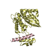
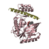
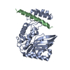
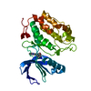
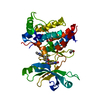
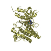
 Homo sapiens (human)
 X-RAY DIFFRACTION /
  Authors
 Citation
 Structure visualization
 Downloads & links
 Other downloads
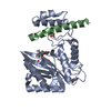

 PDBj
PDBj Assembly
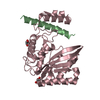

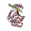
 Spodoptera frugiperda (fall armyworm) / References: UniProt: Q7L8A9, tubulinyl-Tyr carboxypeptidase
 Mass: 92.094 Da / Num. of mol.: 2 / Source method: obtained synthetically / Formula: C3H8O3
 Mass: 18.015 Da / Num. of mol.: 76 / Source method: isolated from a natural source / Formula: H2O
 Sample preparation
 / Beamline: MASSIF-1 / Wavelength: 0.966 Å
 Processing