[English] 日本語
 Yorodumi
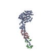
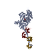
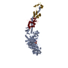
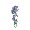
- PDB-2w4w: Isometrically contracting insect asynchronous flight muscle quick... -
+ Open data
- Basic information
| Entry | Database: PDB / ID: 2w4w | ||||||
|---|---|---|---|---|---|---|---|
| Title | Isometrically contracting insect asynchronous flight muscle quick frozen after a quick stretch step | ||||||
 Components Components |
| ||||||
 Keywords Keywords | CONTRACTILE PROTEIN / METHYLATION / ATP-BINDING / ISOMETRIC CONTRACTION / MICROTOMY / FREEZE SUBSTITUTION / MUSCLE PROTEIN / CALMODULIN-BINDING / MOTOR PROTEIN / ACTIN-BINDING | ||||||
| Function / homology |  Function and homology information Function and homology informationmuscle myosin complex / myosin filament / myosin complex / myosin II complex / microfilament motor activity / myofibril / actin filament binding / calmodulin binding / calcium ion binding / ATP binding Similarity search - Function | ||||||
| Biological species |  ARGOPECTEN IRRADIANS (bay scallop) ARGOPECTEN IRRADIANS (bay scallop) | ||||||
| Method | ELECTRON MICROSCOPY / electron tomography / Resolution: 35 Å | ||||||
 Authors Authors | Wu, S. / Liu, J. / Reedy, M.C. / Tregear, R.T. / Winkler, H. / Franzini-Armstrong, C. / Sasaki, H. / Lucaveche, C. / Goldman, Y.E. / Reedy, M.K. / Taylor, K.A. | ||||||
 Citation Citation |  Journal: PLoS One / Year: 2012 Journal: PLoS One / Year: 2012Title: Structural changes in isometrically contracting insect flight muscle trapped following a mechanical perturbation. Authors: Shenping Wu / Jun Liu / Mary C Reedy / Robert J Perz-Edwards / Richard T Tregear / Hanspeter Winkler / Clara Franzini-Armstrong / Hiroyuki Sasaki / Carmen Lucaveche / Yale E Goldman / ...Authors: Shenping Wu / Jun Liu / Mary C Reedy / Robert J Perz-Edwards / Richard T Tregear / Hanspeter Winkler / Clara Franzini-Armstrong / Hiroyuki Sasaki / Carmen Lucaveche / Yale E Goldman / Michael K Reedy / Kenneth A Taylor /  Abstract: The application of rapidly applied length steps to actively contracting muscle is a classic method for synchronizing the response of myosin cross-bridges so that the average response of the ensemble ...The application of rapidly applied length steps to actively contracting muscle is a classic method for synchronizing the response of myosin cross-bridges so that the average response of the ensemble can be measured. Alternatively, electron tomography (ET) is a technique that can report the structure of the individual members of the ensemble. We probed the structure of active myosin motors (cross-bridges) by applying 0.5% changes in length (either a stretch or a release) within 2 ms to isometrically contracting insect flight muscle (IFM) fibers followed after 5-6 ms by rapid freezing against a liquid helium cooled copper mirror. ET of freeze-substituted fibers, embedded and thin-sectioned, provides 3-D cross-bridge images, sorted by multivariate data analysis into ~40 classes, distinct in average structure, population size and lattice distribution. Individual actin subunits are resolved facilitating quasi-atomic modeling of each class average to determine its binding strength (weak or strong) to actin. ~98% of strong-binding acto-myosin attachments present after a length perturbation are confined to "target zones" of only two actin subunits located exactly midway between successive troponin complexes along each long-pitch helical repeat of actin. Significant changes in the types, distribution and structure of actin-myosin attachments occurred in a manner consistent with the mechanical transients. Most dramatic is near disappearance, after either length perturbation, of a class of weak-binding cross-bridges, attached within the target zone, that are highly likely to be precursors of strong-binding cross-bridges. These weak-binding cross-bridges were originally observed in isometrically contracting IFM. Their disappearance following a quick stretch or release can be explained by a recent kinetic model for muscle contraction, as behaviour consistent with their identification as precursors of strong-binding cross-bridges. The results provide a detailed model for contraction in IFM that may be applicable to contraction in other types of muscle. #1: Journal: J Struct Biol / Year: 2009 Title: Methods for identifying and averaging variable molecular conformations in tomograms of actively contracting insect flight muscle. Authors: Shenping Wu / Jun Liu / Mary C Reedy / Hanspeter Winkler / Michael K Reedy / Kenneth A Taylor /  Abstract: During active muscle contraction, tension is generated through many simultaneous, independent interactions between the molecular motor myosin and the actin filaments. The ensemble of myosin motors ...During active muscle contraction, tension is generated through many simultaneous, independent interactions between the molecular motor myosin and the actin filaments. The ensemble of myosin motors displays heterogeneous conformations reflecting different mechanochemical steps of the ATPase pathway. We used electron tomography of actively contracting insect flight muscle fast-frozen, freeze substituted, Araldite embedded, thin-sectioned and stained, to obtain 3D snapshots of the multiplicity of actin-attached myosin structures. We describe procedures for alignment of the repeating lattice of sub-volumes (38.7 nm cross-bridge repeats bounded by troponin) and multivariate data analysis to identify self-similar repeats for computing class averages. Improvements in alignment and classification of repeat sub-volumes reveals (for the first time in active muscle images) the helix of actin subunits in the thin filament and the troponin density with sufficient clarity that a quasiatomic model of the thin filament can be built into the class averages independent of the myosin cross-bridges. We show how quasiatomic model building can identify both strong and weak myosin attachments to actin. We evaluate the accuracy of image classification to enumerate the different types of actin-myosin attachments. | ||||||
| History |
|
- Structure visualization
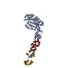
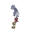
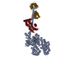
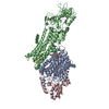
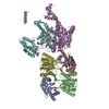
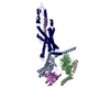
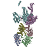
| Movie |
 Movie viewer Movie viewer |
|---|---|
| Structure viewer | Molecule:  Molmil Molmil Jmol/JSmol Jmol/JSmol |
- Downloads & links
- Download
| PDBx/mmCIF format |  2w4w.cif.gz 2w4w.cif.gz | 6.7 MB | Display |  PDBx/mmCIF format PDBx/mmCIF format |
|---|---|---|---|---|
| PDB format |  pdb2w4w.ent.gz pdb2w4w.ent.gz | 5.5 MB | Display |  PDB format PDB format |
| PDBx/mmJSON format |  2w4w.json.gz 2w4w.json.gz | Tree view |  PDBx/mmJSON format PDBx/mmJSON format | |
| Others |  Other downloads Other downloads |
-Validation report
| Summary document |  2w4w_validation.pdf.gz 2w4w_validation.pdf.gz | 1.4 MB | Display |  wwPDB validaton report wwPDB validaton report |
|---|---|---|---|---|
| Full document |  2w4w_full_validation.pdf.gz 2w4w_full_validation.pdf.gz | 12.2 MB | Display | |
| Data in XML |  2w4w_validation.xml.gz 2w4w_validation.xml.gz | 2.7 MB | Display | |
| Data in CIF |  2w4w_validation.cif.gz 2w4w_validation.cif.gz | 3.5 MB | Display | |
| Arichive directory |  https://data.pdbj.org/pub/pdb/validation_reports/w4/2w4w https://data.pdbj.org/pub/pdb/validation_reports/w4/2w4w ftp://data.pdbj.org/pub/pdb/validation_reports/w4/2w4w ftp://data.pdbj.org/pub/pdb/validation_reports/w4/2w4w | HTTPS FTP |
-Related structure data
| Related structure data |  1584MC  1585MC  2w4hC  2w4uC  2w4vC M: map data used to model this data C: citing same article ( |
|---|---|
| Similar structure data |
- Links
Links
- Assembly
| Deposited unit | 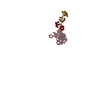
|
|---|---|
| 1 |
|
| Number of models | 40 |
- Components
| #1: Protein | Mass: 94843.883 Da / Num. of mol.: 1 / Fragment: RESIDUES 5-835 / Source method: isolated from a natural source / Details: MYOSIN S1 HEAVY CHAIN / Source: (natural)  ARGOPECTEN IRRADIANS (bay scallop) / References: UniProt: P24733 ARGOPECTEN IRRADIANS (bay scallop) / References: UniProt: P24733 |
|---|---|
| #2: Protein | Mass: 15575.700 Da / Num. of mol.: 1 / Fragment: RESIDUES 16-151 / Source method: isolated from a natural source Details: MYOSIN REGULATORY LIGHT CHAIN, RLC, STRIATED ADDUCTOR MUSCLE Source: (natural)  ARGOPECTEN IRRADIANS (bay scallop) / References: UniProt: P13543 ARGOPECTEN IRRADIANS (bay scallop) / References: UniProt: P13543 |
| #3: Protein | Mass: 17051.912 Da / Num. of mol.: 1 / Fragment: RESIDUES 5-155 / Source method: isolated from a natural source Details: MYOSIN ESSENTIAL LIGHT CHAIN, ELC, STRIATED ADDUCTOR MUSCLE Source: (natural)  ARGOPECTEN IRRADIANS (bay scallop) / References: UniProt: P07291 ARGOPECTEN IRRADIANS (bay scallop) / References: UniProt: P07291 |
| Sequence details | 92 IDENTITY TO P24733 100 IDENTITY TO P13543 100 IDENTITY TO P07291 |
-Experimental details
-Experiment
| Experiment | Method: ELECTRON MICROSCOPY |
|---|---|
| EM experiment | Aggregation state: TISSUE / 3D reconstruction method: electron tomography |
- Sample preparation
| Component | Name: INSECT FIBRILLAR FLIGHT MUSCLE / Type: TISSUE Details: THIS SPECIMEN IS OBTAINED FROM A QUICK FROZEN, ISOMETRICALLY CONTRACTING ASYNCHRONOUS INSECT FLIGHT MUSCLE THAT HAS BEEN FREEZE SUBSTITUTED, PLASTIC EMBEDDED, AND THIN SECTIONED. THE FIBERS ...Details: THIS SPECIMEN IS OBTAINED FROM A QUICK FROZEN, ISOMETRICALLY CONTRACTING ASYNCHRONOUS INSECT FLIGHT MUSCLE THAT HAS BEEN FREEZE SUBSTITUTED, PLASTIC EMBEDDED, AND THIN SECTIONED. THE FIBERS FOR THIS STUDY WERE SUBJECTED TO MECHANICAL TRANSIENTS. FOR THE QUICK RELEASE,THE FIBER WAS RELEASED 9 NM PER HALF-SARCOMERE IN 2 MS AND THE FREEZING IMPACT OCC-6 MS LATER. DATA AQUISITION |
|---|---|
| Buffer solution | Name: 20 MM MOPS BUFFER, 5 MM NAN3, AND MGCL2, ATP, CACL2, AND EGTA IN VARYING MILLIMOLAR CONCENTRATIONS Details: 20 MM MOPS BUFFER, 5 MM NAN3, AND MGCL2, ATP, CACL2, AND EGTA IN VARYING MILLIMOLAR CONCENTRATIONS |
| Specimen | Embedding applied: YES / Shadowing applied: NO / Staining applied: NO / Vitrification applied: NO |
| Specimen support | Details: CARBON |
| Vitrification | Instrument: HOMEMADE PLUNGER / Cryogen name: HELIUM Details: SMASH AGAINST A LIQUID HELIUM COOLED GOLD COATED COPPER MIRROR |
- Electron microscopy imaging
| Microscopy | Model: FEI/PHILIPS CM300FEG/T |
|---|---|
| Electron gun | Electron source:  FIELD EMISSION GUN / Accelerating voltage: 300 kV / Illumination mode: FLOOD BEAM FIELD EMISSION GUN / Accelerating voltage: 300 kV / Illumination mode: FLOOD BEAM |
| Electron lens | Mode: BRIGHT FIELD / Cs: 2 mm |
| Specimen holder | Tilt angle max: 72 ° / Tilt angle min: -72 ° |
| Image recording | Film or detector model: TVIPS TEMCAM-F224 (2k x 2k) |
| Radiation wavelength | Relative weight: 1 |
- Processing
| Symmetry | Point symmetry: C1 (asymmetric) | ||||||||||||
|---|---|---|---|---|---|---|---|---|---|---|---|---|---|
| 3D reconstruction | Resolution method: FSC 0.5 CUT-OFF Details: NOTE THAT OUR LOWEST RESOLUTION DATA IS AT INVERSE 1 MICRON. NUMBER OF FOURIER COEFFICIENTS IS ALMOST A HALF MILLION. THESE COORDINATES WERE FITTED TO AVERAGED SUBVOLUMES OBTAINED FROM A ...Details: NOTE THAT OUR LOWEST RESOLUTION DATA IS AT INVERSE 1 MICRON. NUMBER OF FOURIER COEFFICIENTS IS ALMOST A HALF MILLION. THESE COORDINATES WERE FITTED TO AVERAGED SUBVOLUMES OBTAINED FROM A DUAL AXIS TOMOGRAM. THE FITTING WAS DONE MANUALLY USING THE CRYSTALLOGRAPHIC MODEL FITTING PROGRAM O. THERE ARE 24 MODELS. Symmetry type: POINT | ||||||||||||
| Refinement | Highest resolution: 35 Å | ||||||||||||
| Refinement step | Cycle: LAST / Highest resolution: 35 Å
|
 Movie
Movie Controller


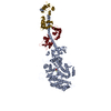
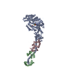
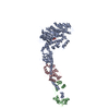
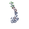



 PDBj
PDBj


