 Movie
Movie Controller


+ Open data

- Basic information
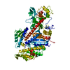
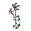
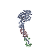
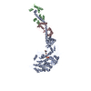
| Entry | Database: PDB / ID: 1b7t | ||||||
|---|---|---|---|---|---|---|---|
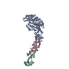
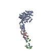
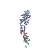
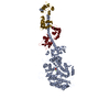
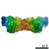
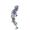
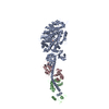
| Title | MYOSIN DIGESTED BY PAPAIN | ||||||
 Components Components |
| ||||||
 Keywords Keywords | MYOSIN / MYOSIN MOTOR | ||||||
| Function / homology |  Function and homology information Function and homology informationmyosin filament organization / myofibril assembly / muscle myosin complex / myosin filament / myosin complex / locomotion / myosin II complex / structural constituent of muscle / microfilament motor activity / myofibril ...myosin filament organization / myofibril assembly / muscle myosin complex / myosin filament / myosin complex / locomotion / myosin II complex / structural constituent of muscle / microfilament motor activity / myofibril / muscle contraction / actin filament binding / calmodulin binding / calcium ion binding / ATP binding Similarity search - Function | ||||||
| Biological species |  Argopecten irradians (bay scallop) Argopecten irradians (bay scallop) | ||||||
| Method |  X-RAY DIFFRACTION / SYNCHROTRON / MOLECULAR REPLACEMENT / Resolution: 2.5 Å X-RAY DIFFRACTION / SYNCHROTRON / MOLECULAR REPLACEMENT / Resolution: 2.5 Å | ||||||
 Authors Authors | Houdusse, A. / Kalabokis, V. / Himmel, D. / Szent-Gyorgyi, A.G. / Cohen, C. | ||||||
 Citation Citation | Journal: Cell / Year: 1999 Title: Atomic structure of scallop myosin subfragment S1 complexed with MgADP: a novel conformation of the myosin head. Authors: A Houdusse / V N Kalabokis / D Himmel / A G Szent-Györgyi / C Cohen /  Abstract: The crystal structure of a proteolytic subfragment from scallop striated muscle myosin, complexed with MgADP, has been solved at 2.5 A resolution and reveals an unusual conformation of the myosin ...The crystal structure of a proteolytic subfragment from scallop striated muscle myosin, complexed with MgADP, has been solved at 2.5 A resolution and reveals an unusual conformation of the myosin head. The converter and the lever arm are in very different positions from those in either the pre-power stroke or near-rigor state structures; moreover, in contrast to these structures, the SH1 helix is seen to be unwound. Here we compare the overall organization of the myosin head in these three states and show how the conformation of three flexible "joints" produces rearrangements of the four major subdomains in the myosin head with different bound nucleotides. We believe that this novel structure represents one of the prehydrolysis ("ATP") states of the contractile cycle in which the myosin heads stay detached from actin. | ||||||
| History |
|
- Structure visualization
| Structure viewer | Molecule: MolmilJmol/JSmol |
|---|
- Downloads & links
-Download
| PDBx/mmCIF format | 1b7t.cif.gz | 226.4 KB | Display | PDBx/mmCIF format |
|---|---|---|---|---|
| PDB format | pdb1b7t.ent.gz | 174.8 KB | Display | PDB format |
| PDBx/mmJSON format | 1b7t.json.gz | Tree view | PDBx/mmJSON format | |
| Others |  Other downloads Other downloads |
-Validation report
| Arichive directory | https://data.pdbj.org/pub/pdb/validation_reports/b7/1b7tftp://data.pdbj.org/pub/pdb/validation_reports/b7/1b7t | HTTPS FTP |
|---|
-Related structure data

-Links
 PDBj
PDBj


- Assembly
| Deposited unit | 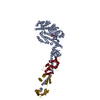
| ||||||||||
|---|---|---|---|---|---|---|---|---|---|---|---|
| 1 |
| ||||||||||
| Unit cell |
|
-Components
-Protein , 3 types, 3 molecules AYZ
| #1: Protein | Mass: 95317.430 Da / Num. of mol.: 1 / Fragment: PAPAIN DIGESTED, SUBFRAGMENT 1 (S1) / Source method: isolated from a natural source / Details: PAPAIN DIGESTION OF MYOSIN / Source: (natural) Argopecten irradians (bay scallop) / Tissue: SKELETAL MUSCLE / References: UniProt: P24733 |
|---|---|
| #2: Protein | Mass: 17560.855 Da / Num. of mol.: 1 / Fragment: PAPAIN DIGESTED, SUBFRAGMENT 1 (S1) / Source method: isolated from a natural source / Details: PAPAIN DIGESTION OF MYOSIN / Source: (natural) Argopecten irradians (bay scallop) / Tissue: SKELETAL MUSCLE / References: UniProt: P13543 |
| #3: Protein | Mass: 17635.635 Da / Num. of mol.: 1 / Fragment: PAPAIN DIGESTED, SUBFRAGMENT 1 (S1) / Source method: isolated from a natural source / Details: PAPAIN DIGESTION OF MYOSIN / Source: (natural) Argopecten irradians (bay scallop) / Tissue: SKELETAL MUSCLE / References: UniProt: P07291 |
-Non-polymers , 4 types, 108 molecules 


| #4: Chemical |  Mass: 24.305 Da / Num. of mol.: 2 / Source method: obtained synthetically / Formula: Mg Mass: 24.305 Da / Num. of mol.: 2 / Source method: obtained synthetically / Formula: Mg#5: Chemical | ChemComp-ADP / |  Mass: 427.201 Da / Num. of mol.: 1 / Source method: obtained synthetically / Formula: C10H15N5O10P2 / Comment: ADP, energy-carrying molecule*YM Mass: 427.201 Da / Num. of mol.: 1 / Source method: obtained synthetically / Formula: C10H15N5O10P2 / Comment: ADP, energy-carrying molecule*YM#6: Chemical | ChemComp-CA / |  Mass: 40.078 Da / Num. of mol.: 1 / Source method: obtained synthetically / Formula: Ca Mass: 40.078 Da / Num. of mol.: 1 / Source method: obtained synthetically / Formula: Ca#7: Water | ChemComp-HOH / | Mass: 18.015 Da / Num. of mol.: 104 / Source method: isolated from a natural source / Formula: H2O |
|---|
-Experimental details
-Experiment
| Experiment | Method: X-RAY DIFFRACTION / Number of used crystals: 1 |
|---|
- Sample preparation
| Crystal | Density Matthews: 3.11 Å3/Da / Density % sol: 64 % | ||||||||||||||||||||||||||||||||||||||||||
|---|---|---|---|---|---|---|---|---|---|---|---|---|---|---|---|---|---|---|---|---|---|---|---|---|---|---|---|---|---|---|---|---|---|---|---|---|---|---|---|---|---|---|---|
| Crystal grow | pH: 8 / Details: pH 8.0 | ||||||||||||||||||||||||||||||||||||||||||
| Crystal | *PLUS Density % sol: 64.6 % | ||||||||||||||||||||||||||||||||||||||||||
| Crystal grow | *PLUS Temperature: 4 ℃ / Method: vapor diffusion, sitting drop / Details: used to seeding | ||||||||||||||||||||||||||||||||||||||||||
| Components of the solutions | *PLUS
|
-Data collection
| Diffraction | Mean temperature: 100 K |
|---|---|
| Diffraction source | Source: SYNCHROTRON / Site: CHESS / Beamline: F1 / Wavelength: 0.918 |
| Detector | Detector: CCD / Date: Jan 15, 1997 |
| Radiation | Protocol: SINGLE WAVELENGTH / Monochromatic (M) / Laue (L): M / Scattering type: x-ray |
| Radiation wavelength | Wavelength: 0.918 Å / Relative weight: 1 |
| Reflection | Resolution: 2.5→40 Å / Num. obs: 43838 / % possible obs: 80.6 % / Redundancy: 3 % / Biso Wilson estimate: 50 Å2 / Rmerge(I) obs: 0.048 / Rsym value: 0.048 / Net I/σ(I): 11.6 |
| Reflection shell | *PLUS Highest resolution: 2.5 Å / Lowest resolution: 2.8 Å / % possible obs: 38.5 % / Redundancy: 1.4 % / Num. unique obs: 5968 / Rmerge(I) obs: 0.11 / Mean I/σ(I) obs: 6 |
- Processing
| Software |
| ||||||||||||||||||||||||||||||||||||||||||||||||||||||||||||
|---|---|---|---|---|---|---|---|---|---|---|---|---|---|---|---|---|---|---|---|---|---|---|---|---|---|---|---|---|---|---|---|---|---|---|---|---|---|---|---|---|---|---|---|---|---|---|---|---|---|---|---|---|---|---|---|---|---|---|---|---|---|
| Refinement | Method to determine structure: MOLECULAR REPLACEMENT Starting model: 1MMD, 1WDC Resolution: 2.5→30 Å / Isotropic thermal model: RESTRAINED / Cross valid method: THROUGHOUT / σ(F): 0
| ||||||||||||||||||||||||||||||||||||||||||||||||||||||||||||
| Refine analyze |
| ||||||||||||||||||||||||||||||||||||||||||||||||||||||||||||
| Refinement step | Cycle: LAST / Resolution: 2.5→30 Å
| ||||||||||||||||||||||||||||||||||||||||||||||||||||||||||||
| Refine LS restraints |
| ||||||||||||||||||||||||||||||||||||||||||||||||||||||||||||
| Xplor file | Serial no: 1 / Param file: PROTEIN_REP.PARAM / Topol file: TOPHCSDX.PRO | ||||||||||||||||||||||||||||||||||||||||||||||||||||||||||||
| Software | *PLUS Name: X-PLOR / Version: 4 / Classification: refinement | ||||||||||||||||||||||||||||||||||||||||||||||||||||||||||||
| Refinement | *PLUS Highest resolution: 2.5 Å / Lowest resolution: 30 Å / σ(F): 0 / % reflection Rfree: 5 % / Rfactor obs: 0.2244 | ||||||||||||||||||||||||||||||||||||||||||||||||||||||||||||
| Solvent computation | *PLUS | ||||||||||||||||||||||||||||||||||||||||||||||||||||||||||||
| Displacement parameters | *PLUS | ||||||||||||||||||||||||||||||||||||||||||||||||||||||||||||
| Refine LS restraints | *PLUS
|
