 Movie
Movie Controller

[English] 日本語
 Yorodumi
- PDB-2os8: Rigor-like structures of muscle myosins reveal key mechanical ele... -
+ Open data

- Basic information
| Entry | Database: PDB / ID: 2os8 | ||||||
|---|---|---|---|---|---|---|---|
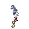
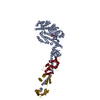
| Title | Rigor-like structures of muscle myosins reveal key mechanical elements in the transduction pathways of this allosteric motor | ||||||
 Components Components |
| ||||||
 Keywords Keywords | CONTRACTILE PROTEIN / myosin S1 / rigor-like / motor / mechanical elements | ||||||
| Function / homology |  Function and homology information Function and homology informationmyosin filament organization / myofibril assembly / muscle myosin complex / myosin filament / locomotion / myosin complex / myosin II complex / structural constituent of muscle / microfilament motor activity / myofibril ...myosin filament organization / myofibril assembly / muscle myosin complex / myosin filament / locomotion / myosin complex / myosin II complex / structural constituent of muscle / microfilament motor activity / myofibril / muscle contraction / actin filament binding / calcium ion binding / ATP binding / metal ion binding Similarity search - Function | ||||||
| Biological species |  Placopecten magellanicus (sea scallop) Placopecten magellanicus (sea scallop) | ||||||
| Method |  X-RAY DIFFRACTION / SYNCHROTRON / MOLECULAR REPLACEMENT / Resolution: 3.27 Å X-RAY DIFFRACTION / SYNCHROTRON / MOLECULAR REPLACEMENT / Resolution: 3.27 Å | ||||||
 Authors Authors | Yang, Y. / Gourinath, S. / Cohen, C. / Brown, J.H. | ||||||
 Citation Citation | Journal: Structure / Year: 2007 Title: Rigor-like Structures from Muscle Myosins Reveal Key Mechanical Elements in the Transduction Pathways of This Allosteric Motor. Authors: Yang, Y. / Gourinath, S. / Kovacs, M. / Nyitray, L. / Reutzel, R. / Himmel, D.M. / O'neall-Hennessey, E. / Reshetnikova, L. / Szent-Gyorgyi, A.G. / Brown, J.H. / Cohen, C. | ||||||
| History |
|
- Structure visualization
| Structure viewer | Molecule: MolmilJmol/JSmol |
|---|
- Downloads & links
-Download
| PDBx/mmCIF format | 2os8.cif.gz | 230.3 KB | Display | PDBx/mmCIF format |
|---|---|---|---|---|
| PDB format | pdb2os8.ent.gz | 180.7 KB | Display | PDB format |
| PDBx/mmJSON format | 2os8.json.gz | Tree view | PDBx/mmJSON format | |
| Others |  Other downloads Other downloads |
-Validation report
| Arichive directory | https://data.pdbj.org/pub/pdb/validation_reports/os/2os8ftp://data.pdbj.org/pub/pdb/validation_reports/os/2os8 | HTTPS FTP |
|---|
-Related structure data
| Related structure data |  2ec6C  2otgC  3i5fC  3i5gC  3i5hC  3i5iC  1sr6S S: Starting model for refinement C: citing same article ( |
|---|---|
| Similar structure data |


-Links
 PDBj
PDBj


- Assembly
| Deposited unit | 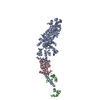
| ||||||||
|---|---|---|---|---|---|---|---|---|---|
| 1 |
| ||||||||
| Unit cell |
|
-Components
| #1: Protein | Mass: 96139.289 Da / Num. of mol.: 1 / Fragment: Myosin heavy chain / Source method: isolated from a natural source / Source: (natural) Placopecten magellanicus (sea scallop) / References: UniProt: Q26080 |
|---|---|
| #2: Protein | Mass: 17769.146 Da / Num. of mol.: 1 / Fragment: Myosin RLC / Source method: isolated from a natural source / Source: (natural) Placopecten magellanicus (sea scallop) / References: UniProt: Q26068 |
| #3: Protein | Mass: 17811.805 Da / Num. of mol.: 1 / Fragment: Myosin ELC / Source method: isolated from a natural source / Source: (natural) Placopecten magellanicus (sea scallop) / References: UniProt: Q26066 |
| #4: Chemical | ChemComp-MG /   Mass: 24.305 Da / Num. of mol.: 1 / Source method: obtained synthetically / Formula: Mg Mass: 24.305 Da / Num. of mol.: 1 / Source method: obtained synthetically / Formula: Mg |
| #5: Chemical | ChemComp-CA /   Mass: 40.078 Da / Num. of mol.: 1 / Source method: obtained synthetically / Formula: Ca Mass: 40.078 Da / Num. of mol.: 1 / Source method: obtained synthetically / Formula: Ca |
-Experimental details
-Experiment
| Experiment | Method: X-RAY DIFFRACTION / Number of used crystals: 2 |
|---|
- Sample preparation
| Crystal | Density Matthews: 2.61 Å3/Da / Density % sol: 52.91 % |
|---|---|
| Crystal grow | Temperature: 277 K / Method: vapor diffusion, hanging drop / pH: 7.5 Details: CaCl2, PEG , pH 7.5, VAPOR DIFFUSION, HANGING DROP, temperature 277K |
-Data collection
| Diffraction | Mean temperature: 100 K |
|---|---|
| Diffraction source | Source: SYNCHROTRON / Site: CHESS  / Beamline: A1 / Wavelength: 0.98 Å / Beamline: A1 / Wavelength: 0.98 Å |
| Detector | Type: ADSC QUANTUM 4 / Detector: CCD |
| Radiation | Protocol: SINGLE WAVELENGTH / Monochromatic (M) / Laue (L): M / Scattering type: x-ray |
| Radiation wavelength | Wavelength: 0.98 Å / Relative weight: 1 |
| Reflection | Resolution: 3.25→50 Å / Num. all: 16888 / Num. obs: 16888 / % possible obs: 79.6 % / Observed criterion σ(I): -3 / Redundancy: 3.5 % / Biso Wilson estimate: 66 Å2 / Rsym value: 0.085 / Net I/σ(I): 7.2 |
| Reflection shell | Resolution: 3.25→3.37 Å / Redundancy: 2.5 % / Mean I/σ(I) obs: 1.5 / Num. unique all: 1371 / Rsym value: 0.451 / % possible all: 64.7 |
- Processing
| Software |
| ||||||||||||||||||||||||||||
|---|---|---|---|---|---|---|---|---|---|---|---|---|---|---|---|---|---|---|---|---|---|---|---|---|---|---|---|---|---|
| Refinement | Method to determine structure: MOLECULAR REPLACEMENT Starting model: 1SR6 Resolution: 3.27→48.03 Å / Rfactor Rfree error: 0.009 / Data cutoff high absF: 125059.531 / Data cutoff low absF: 0 / Isotropic thermal model: RESTRAINED / Cross valid method: THROUGHOUT / σ(F): 0 / Stereochemistry target values: Engh & Huber
| ||||||||||||||||||||||||||||
| Solvent computation | Solvent model: FLAT MODEL / Bsol: 117.409 Å2 / ksol: 0.261 e/Å3 | ||||||||||||||||||||||||||||
| Displacement parameters | Biso mean: 102.2 Å2
| ||||||||||||||||||||||||||||
| Refine analyze |
| ||||||||||||||||||||||||||||
| Refinement step | Cycle: LAST / Resolution: 3.27→48.03 Å
| ||||||||||||||||||||||||||||
| Refine LS restraints |
| ||||||||||||||||||||||||||||
| LS refinement shell | Resolution: 3.25→3.45 Å / Rfactor Rfree error: 0.033 / Total num. of bins used: 6
| ||||||||||||||||||||||||||||
| Xplor file |
|
