 Movie
Movie Controller

[English] 日本語
 Yorodumi
- PDB-1s5g: Structure of Scallop myosin S1 reveals a novel nucleotide conformation -
+ Open data

- Basic information
| Entry | Database: PDB / ID: 1s5g | ||||||
|---|---|---|---|---|---|---|---|
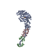
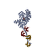
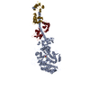
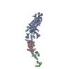
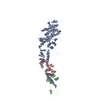
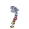
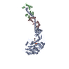
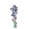
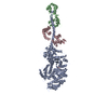
| Title | Structure of Scallop myosin S1 reveals a novel nucleotide conformation | ||||||
 Components Components |
| ||||||
 Keywords Keywords | CONTRACTILE PROTEIN / Scallop myosin S1 / Near Rigor / complex salt bridge / novel conformation of nucleotide | ||||||
| Function / homology |  Function and homology information Function and homology informationmyosin filament organization / myofibril assembly / muscle myosin complex / myosin filament / myosin complex / locomotion / myosin II complex / structural constituent of muscle / microfilament motor activity / myofibril ...myosin filament organization / myofibril assembly / muscle myosin complex / myosin filament / myosin complex / locomotion / myosin II complex / structural constituent of muscle / microfilament motor activity / myofibril / muscle contraction / actin filament binding / calmodulin binding / calcium ion binding / ATP binding Similarity search - Function | ||||||
| Biological species |  Argopecten irradians (bay scallop) Argopecten irradians (bay scallop) | ||||||
| Method |  X-RAY DIFFRACTION / SYNCHROTRON / MOLECULAR REPLACEMENT / Resolution: 3.1 Å X-RAY DIFFRACTION / SYNCHROTRON / MOLECULAR REPLACEMENT / Resolution: 3.1 Å | ||||||
 Authors Authors | Risal, D. / Gourinath, S. / Himmel, D.M. / Szent-Gyorgyi, A.G. / Cohen, C. | ||||||
 Citation Citation | Journal: Proc.Natl.Acad.Sci.Usa / Year: 2004 Title: Myosin subfragment 1 structures reveal a partially bound nucleotide and a complex salt bridge that helps couple nucleotide and actin binding. Authors: Risal, D. / Gourinath, S. / Himmel, D.M. / Szent-Gyorgyi, A.G. / Cohen, C. #1: Journal: Structure / Year: 2003Title: Crystal Structure Of Scallop Myosin S1 In The Pre-Power Stroke State To 2.6 A Resolution: Flexibility and Function In The Head Authors: Gourinath, S. / Himmel, D.M. / Brown, J.H. / Reshetnikova, L. / Szent-Gyorgyi, A.G. / Cohen, C. #2: Journal: Proc.Natl.Acad.Sci.USA / Year: 2002Title: Crystallographic findings on the internally uncoupled and near-rigor states of myosin: further insights into the mechanics of the motor Authors: Himmel, D.M. / Gourinath, S. / Reshetnikova, L. / Shen, Y. / Szent-Gyorgyi, A.G. / Cohen, C. | ||||||
| History |
|
- Structure visualization
| Structure viewer | Molecule: MolmilJmol/JSmol |
|---|
- Downloads & links
-Download
| PDBx/mmCIF format | 1s5g.cif.gz | 231.9 KB | Display | PDBx/mmCIF format |
|---|---|---|---|---|
| PDB format | pdb1s5g.ent.gz | 181 KB | Display | PDB format |
| PDBx/mmJSON format | 1s5g.json.gz | Tree view | PDBx/mmJSON format | |
| Others |  Other downloads Other downloads |
-Validation report
| Arichive directory | https://data.pdbj.org/pub/pdb/validation_reports/s5/1s5gftp://data.pdbj.org/pub/pdb/validation_reports/s5/1s5g | HTTPS FTP |
|---|
-Related structure data
| Related structure data | 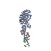 1sr6C  1kk7S S: Starting model for refinement C: citing same article ( |
|---|---|
| Similar structure data |


-Links
 PDBj
PDBj


- Assembly
| Deposited unit | 
| ||||||||
|---|---|---|---|---|---|---|---|---|---|
| 1 |
| ||||||||
| Unit cell |
|
-Components
-Protein , 3 types, 3 molecules AYZ
| #1: Protein | Mass: 95815.062 Da / Num. of mol.: 1 / Fragment: Residues 1-840 / Source method: isolated from a natural source / Source: (natural) Argopecten irradians (bay scallop) / References: UniProt: P24733 |
|---|---|
| #2: Protein | Mass: 17560.855 Da / Num. of mol.: 1 / Source method: isolated from a natural source / Source: (natural) Argopecten irradians (bay scallop) / References: UniProt: P13543 |
| #3: Protein | Mass: 17635.635 Da / Num. of mol.: 1 / Source method: isolated from a natural source / Source: (natural) Argopecten irradians (bay scallop) / References: UniProt: P07291 |
-Non-polymers , 5 types, 5 molecules 


| #4: Chemical | ChemComp-SO4 /  Mass: 96.063 Da / Num. of mol.: 1 / Source method: obtained synthetically / Formula: SO4 Mass: 96.063 Da / Num. of mol.: 1 / Source method: obtained synthetically / Formula: SO4 |
|---|---|
| #5: Chemical | ChemComp-ADP /  Mass: 427.201 Da / Num. of mol.: 1 / Source method: obtained synthetically / Formula: C10H15N5O10P2 / Comment: ADP, energy-carrying molecule*YM Mass: 427.201 Da / Num. of mol.: 1 / Source method: obtained synthetically / Formula: C10H15N5O10P2 / Comment: ADP, energy-carrying molecule*YM |
| #6: Chemical | ChemComp-MG /  Mass: 24.305 Da / Num. of mol.: 1 / Source method: obtained synthetically / Formula: Mg Mass: 24.305 Da / Num. of mol.: 1 / Source method: obtained synthetically / Formula: Mg |
| #7: Chemical | ChemComp-CA /  Mass: 40.078 Da / Num. of mol.: 1 / Source method: obtained synthetically / Formula: Ca Mass: 40.078 Da / Num. of mol.: 1 / Source method: obtained synthetically / Formula: Ca |
| #8: Water | ChemComp-HOH / Mass: 18.015 Da / Num. of mol.: 1 / Source method: isolated from a natural source / Formula: H2O |
-Experimental details
-Experiment
| Experiment | Method: X-RAY DIFFRACTION / Number of used crystals: 3 |
|---|
- Sample preparation
| Crystal | Density Matthews: 2.61 Å3/Da / Density % sol: 52.9 % |
|---|---|
| Crystal grow | Temperature: 300 K / Method: vapor diffusion, sitting drop / pH: 6.5 Details: PEG 8000, magnesium chloride, ADP, Glycerol, MES, TMAO, cacodylate, pH 6.5, VAPOR DIFFUSION, SITTING DROP, temperature 300K |
-Data collection
| Diffraction | Mean temperature: 100 K |
|---|---|
| Diffraction source | Source: SYNCHROTRON / Site: CHESS  / Beamline: A1 / Wavelength: 0.9 Å / Beamline: A1 / Wavelength: 0.9 Å |
| Detector | Type: ADSC QUANTUM 4 / Detector: CCD / Date: Nov 24, 2002 |
| Radiation | Monochromator: RH-COATED WITH SI / Protocol: SINGLE WAVELENGTH / Monochromatic (M) / Laue (L): M / Scattering type: x-ray |
| Radiation wavelength | Wavelength: 0.9 Å / Relative weight: 1 |
| Reflection | Resolution: 3.1→50 Å / Num. all: 22278 / Num. obs: 21309 / % possible obs: 85.2 % / Observed criterion σ(F): -3 / Observed criterion σ(I): 0 / Redundancy: 4.2 % / Biso Wilson estimate: 80.5 Å2 / Rsym value: 0.071 / Net I/σ(I): 20.6 |
| Reflection shell | Resolution: 3.1→3.21 Å / Redundancy: 1.5 % / Mean I/σ(I) obs: 4.2 / Num. unique all: 1553 / Rsym value: 0.284 / % possible all: 59.1 |
- Processing
| Software |
| |||||||||||||||||||||||||
|---|---|---|---|---|---|---|---|---|---|---|---|---|---|---|---|---|---|---|---|---|---|---|---|---|---|---|
| Refinement | Method to determine structure: MOLECULAR REPLACEMENT Starting model: PDB ENTRY 1kk7 Resolution: 3.1→43.44 Å / Isotropic thermal model: OVERALL ANISOTROPIC B VALUE / Cross valid method: THROUGHOUT / σ(F): 0 / Stereochemistry target values: Engh & Huber
| |||||||||||||||||||||||||
| Displacement parameters | Biso mean: 76.4 Å2
| |||||||||||||||||||||||||
| Refine analyze |
| |||||||||||||||||||||||||
| Refinement step | Cycle: LAST / Resolution: 3.1→43.44 Å
| |||||||||||||||||||||||||
| Refine LS restraints |
|
