 Movie
Movie Controller

[English] 日本語
 Yorodumi
- PDB-1qvi: Crystal structure of scallop myosin S1 in the pre-power stroke st... -
+ Open data

- Basic information
| Entry | Database: PDB / ID: 1qvi | ||||||
|---|---|---|---|---|---|---|---|
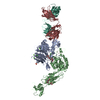
| Title | Crystal structure of scallop myosin S1 in the pre-power stroke state to 2.6 Angstrom resolution: flexibility and function in the head | ||||||
 Components Components |
| ||||||
 Keywords Keywords | CONTRACTILE PROTEIN / scallop myosin subfragment-1 (S1) / pre-power stroke state / pliant region / internally-uncoupled state / SH1 helix | ||||||
| Function / homology |  Function and homology information Function and homology informationmyosin filament organization / myofibril assembly / muscle myosin complex / myosin filament / locomotion / myosin complex / myosin II complex / structural constituent of muscle / microfilament motor activity / myofibril ...myosin filament organization / myofibril assembly / muscle myosin complex / myosin filament / locomotion / myosin complex / myosin II complex / structural constituent of muscle / microfilament motor activity / myofibril / muscle contraction / actin filament binding / calmodulin binding / calcium ion binding / ATP binding Similarity search - Function | ||||||
| Biological species |  Argopecten irradians (bay scallop) Argopecten irradians (bay scallop) | ||||||
| Method |  X-RAY DIFFRACTION / SYNCHROTRON / MOLECULAR REPLACEMENT / Resolution: 2.54 Å X-RAY DIFFRACTION / SYNCHROTRON / MOLECULAR REPLACEMENT / Resolution: 2.54 Å | ||||||
 Authors Authors | Gourinath, S. / Himmel, D.M. / Brown, J.H. / Reshetnikova, L. / Szent-Gyrgyi, A.G. / Cohen, C. | ||||||
 Citation Citation | Journal: Structure / Year: 2003 Title: Crystal structure of scallop Myosin s1 in the pre-power stroke state to 2.6 a resolution: flexibility and function in the head. Authors: Gourinath, S. / Himmel, D.M. / Brown, J.H. / Reshetnikova, L. / Szent-Gyorgyi, A.G. / Cohen, C. | ||||||
| History |
|
- Structure visualization
| Structure viewer | Molecule: MolmilJmol/JSmol |
|---|
- Downloads & links
-Download
| PDBx/mmCIF format | 1qvi.cif.gz | 235.5 KB | Display | PDBx/mmCIF format |
|---|---|---|---|---|
| PDB format | pdb1qvi.ent.gz | 184.8 KB | Display | PDB format |
| PDBx/mmJSON format | 1qvi.json.gz | Tree view | PDBx/mmJSON format | |
| Others |  Other downloads Other downloads |
-Validation report
| Arichive directory | https://data.pdbj.org/pub/pdb/validation_reports/qv/1qviftp://data.pdbj.org/pub/pdb/validation_reports/qv/1qvi | HTTPS FTP |
|---|
-Related structure data
| Related structure data |  1dflS S: Starting model for refinement |
|---|---|
| Similar structure data |


-Links
 PDBj
PDBj


- Assembly
| Deposited unit | 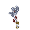
| ||||||||
|---|---|---|---|---|---|---|---|---|---|
| 1 |
| ||||||||
| Unit cell |
|
-Components
-Protein , 3 types, 3 molecules AYZ
| #1: Protein | Mass: 95815.062 Da / Num. of mol.: 1 / Source method: isolated from a natural source / Source: (natural) Argopecten irradians (bay scallop) / References: UniProt: P24733 |
|---|---|
| #2: Protein | Mass: 17560.855 Da / Num. of mol.: 1 / Source method: isolated from a natural source / Source: (natural) Argopecten irradians (bay scallop) / References: UniProt: P13543 |
| #3: Protein | Mass: 17635.635 Da / Num. of mol.: 1 / Source method: isolated from a natural source / Source: (natural) Argopecten irradians (bay scallop) / References: UniProt: P07291 |
-Non-polymers , 5 types, 57 molecules 


| #4: Chemical |  Mass: 24.305 Da / Num. of mol.: 2 / Source method: obtained synthetically / Formula: Mg Mass: 24.305 Da / Num. of mol.: 2 / Source method: obtained synthetically / Formula: Mg#5: Chemical | ChemComp-VO4 / |  Mass: 114.939 Da / Num. of mol.: 1 / Source method: obtained synthetically / Formula: VO4 Mass: 114.939 Da / Num. of mol.: 1 / Source method: obtained synthetically / Formula: VO4#6: Chemical | ChemComp-ADP / |  Mass: 427.201 Da / Num. of mol.: 1 / Source method: obtained synthetically / Formula: C10H15N5O10P2 / Comment: ADP, energy-carrying molecule*YM Mass: 427.201 Da / Num. of mol.: 1 / Source method: obtained synthetically / Formula: C10H15N5O10P2 / Comment: ADP, energy-carrying molecule*YM#7: Chemical | ChemComp-CA / |  Mass: 40.078 Da / Num. of mol.: 1 / Source method: obtained synthetically / Formula: Ca Mass: 40.078 Da / Num. of mol.: 1 / Source method: obtained synthetically / Formula: Ca#8: Water | ChemComp-HOH / | Mass: 18.015 Da / Num. of mol.: 52 / Source method: isolated from a natural source / Formula: H2O |
|---|
-Experimental details
-Experiment
| Experiment | Method: X-RAY DIFFRACTION / Number of used crystals: 3 |
|---|
- Sample preparation
| Crystal | Density Matthews: 3.05 Å3/Da / Density % sol: 59.64 % | ||||||||||||||||||||||||||||||||||||||||||||||||||||||||
|---|---|---|---|---|---|---|---|---|---|---|---|---|---|---|---|---|---|---|---|---|---|---|---|---|---|---|---|---|---|---|---|---|---|---|---|---|---|---|---|---|---|---|---|---|---|---|---|---|---|---|---|---|---|---|---|---|---|
| Crystal grow | Temperature: 298 K / Method: vapor diffusion, sitting drop / pH: 6 Details: MES, MgCl2, ADP, Na3VO4, polyethylene glycol 8K, glycerol, pH 6.0, VAPOR DIFFUSION, SITTING DROP, temperature 298K | ||||||||||||||||||||||||||||||||||||||||||||||||||||||||
| Crystal grow | *PLUS Method: vapor diffusion, sitting drop | ||||||||||||||||||||||||||||||||||||||||||||||||||||||||
| Components of the solutions | *PLUS
|
-Data collection
| Diffraction | Mean temperature: 100 K |
|---|---|
| Diffraction source | Source: SYNCHROTRON / Site: CHESS  / Beamline: A1 / Wavelength: 0.93 Å / Beamline: A1 / Wavelength: 0.93 Å |
| Detector | Type: ADSC QUANTUM 4 / Detector: CCD / Date: Apr 28, 2001 |
| Radiation | Monochromator: RH-COATED WITH SI / Protocol: SINGLE WAVELENGTH / Monochromatic (M) / Laue (L): M / Scattering type: x-ray |
| Radiation wavelength | Wavelength: 0.93 Å / Relative weight: 1 |
| Reflection | Resolution: 2.54→50 Å / Num. all: 42849 / Num. obs: 42805 / % possible obs: 82.3 % / Observed criterion σ(I): -3 / Redundancy: 3.5 % / Biso Wilson estimate: 48.4 Å2 / Rsym value: 0.083 / Net I/σ(I): 18.6 |
| Reflection shell | Resolution: 2.55→2.64 Å / Mean I/σ(I) obs: 5 / Num. unique all: 1696 / Rsym value: 0.18 / % possible all: 33 |
| Reflection | *PLUS Num. obs: 42849 / Rmerge(I) obs: 0.083 |
| Reflection shell | *PLUS % possible obs: 32.8 % / Rmerge(I) obs: 0.18 |
- Processing
| Software |
| |||||||||||||||||||||||||
|---|---|---|---|---|---|---|---|---|---|---|---|---|---|---|---|---|---|---|---|---|---|---|---|---|---|---|
| Refinement | Method to determine structure: MOLECULAR REPLACEMENT Starting model: 1DFL Resolution: 2.54→45.58 Å / Rfactor Rfree error: 0.005 / Isotropic thermal model: RESTRAINED / Cross valid method: THROUGHOUT / σ(F): 0 / Stereochemistry target values: Engh & Huber
| |||||||||||||||||||||||||
| Solvent computation | Solvent model: FLAT MODEL / Bsol: 28.0872 Å2 / ksol: 0.311881 e/Å3 | |||||||||||||||||||||||||
| Displacement parameters | Biso mean: 50.9 Å2
| |||||||||||||||||||||||||
| Refine analyze |
| |||||||||||||||||||||||||
| Refinement step | Cycle: LAST / Resolution: 2.54→45.58 Å
| |||||||||||||||||||||||||
| Refine LS restraints |
| |||||||||||||||||||||||||
| LS refinement shell | Resolution: 2.5→2.66 Å / Rfactor Rfree error: 0.032 / Total num. of bins used: 6
| |||||||||||||||||||||||||
| Xplor file |
| |||||||||||||||||||||||||
| Refinement | *PLUS Lowest resolution: 50 Å / % reflection Rfree: 5 % / Rfactor Rwork: 0.211 | |||||||||||||||||||||||||
| Solvent computation | *PLUS | |||||||||||||||||||||||||
| Displacement parameters | *PLUS | |||||||||||||||||||||||||
| Refine LS restraints | *PLUS
| |||||||||||||||||||||||||
| LS refinement shell | *PLUS % reflection Rfree: 10 % |
