 Movie
Movie Controller

[English] 日本語
 Yorodumi
- PDB-2w2h: Structural basis of transcription activation by the Cyclin T1-Tat... -
+ Open data

- Basic information
| Entry | Database: PDB / ID: 2w2h | ||||||
|---|---|---|---|---|---|---|---|
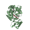
| Title | Structural basis of transcription activation by the Cyclin T1-Tat-TAR RNA complex from EIAV | ||||||
 Components Components |
| ||||||
 Keywords Keywords | RNA BINDING PROTEIN / RNA-BINDING PROTEIN / TAR / TAT / CYCLIN / NUCLEUS / ACTIVATOR / CYCLIN T1 / HOST-VIRUS INTERACTION / TRANSCRIPTION REGULATION / CELL CYCLE / RNA-BINDING / ACETYLATION / COILED COIL / TRANSCRIPTION / CELL DIVISION / CRYSTALLIZATION | ||||||
| Function / homology |  Function and homology information Function and homology informationP-TEFb complex / 7SK snRNA binding / positive regulation of viral transcription / cyclin/CDK positive transcription elongation factor complex / host cell nucleolus / cyclin-dependent protein serine/threonine kinase activator activity / positive regulation of DNA-templated transcription, elongation / RNA-binding transcription regulator activity / positive regulation of transcription elongation by RNA polymerase II / molecular condensate scaffold activity ...P-TEFb complex / 7SK snRNA binding / positive regulation of viral transcription / cyclin/CDK positive transcription elongation factor complex / host cell nucleolus / cyclin-dependent protein serine/threonine kinase activator activity / positive regulation of DNA-templated transcription, elongation / RNA-binding transcription regulator activity / positive regulation of transcription elongation by RNA polymerase II / molecular condensate scaffold activity / cell division / positive regulation of transcription by RNA polymerase II / RNA binding / nucleus Similarity search - Function | ||||||
| Biological species |   EQUINE INFECTIOUS ANEMIA VIRUS EQUINE INFECTIOUS ANEMIA VIRUS | ||||||
| Method |  X-RAY DIFFRACTION / SYNCHROTRON / MOLECULAR REPLACEMENT / Resolution: 3.25 Å X-RAY DIFFRACTION / SYNCHROTRON / MOLECULAR REPLACEMENT / Resolution: 3.25 Å | ||||||
 Authors Authors | Anand, K. / Geyer, M. | ||||||
 Citation Citation | Journal: Nat.Struct.Mol.Biol. / Year: 2008 Title: Structural Insights Into the Cyclin T1-Tat-Tar RNA Transcription Activation Complex from Eiav. Authors: Anand, K. / Schulte, A. / Vogel-Bachmayr, K. / Scheffzek, K. / Geyer, M. | ||||||
| History |
|
- Structure visualization
| Structure viewer | Molecule: MolmilJmol/JSmol |
|---|
- Downloads & links
-Download
| PDBx/mmCIF format | 2w2h.cif.gz | 306.6 KB | Display | PDBx/mmCIF format |
|---|---|---|---|---|
| PDB format | pdb2w2h.ent.gz | 248.3 KB | Display | PDB format |
| PDBx/mmJSON format | 2w2h.json.gz | Tree view | PDBx/mmJSON format | |
| Others |  Other downloads Other downloads |
-Validation report
| Arichive directory | https://data.pdbj.org/pub/pdb/validation_reports/w2/2w2hftp://data.pdbj.org/pub/pdb/validation_reports/w2/2w2h | HTTPS FTP |
|---|
-Related structure data
| Related structure data |  2pk2S S: Starting model for refinement |
|---|---|
| Similar structure data |


-Links
 PDBj
PDBj


- Assembly
| Deposited unit | 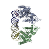
| |||||||||||||||||||||||||||||||||
|---|---|---|---|---|---|---|---|---|---|---|---|---|---|---|---|---|---|---|---|---|---|---|---|---|---|---|---|---|---|---|---|---|---|---|
| 1 | 
| |||||||||||||||||||||||||||||||||
| 2 | 
| |||||||||||||||||||||||||||||||||
| Unit cell |
| |||||||||||||||||||||||||||||||||
| Noncrystallographic symmetry (NCS) | NCS domain:
NCS domain segments:
NCS ensembles :
NCS oper: (Code: given Matrix: (0.0005, -1, 0.0094), Vector: |
-Components
| #1: Protein | Mass: 30843.354 Da / Num. of mol.: 2 / Fragment: RESIDUES 5-267 Source method: isolated from a genetically manipulated source Details: GENBANK\: AF137509 / Source: (gene. exp.)  #2: Protein/peptide | Mass: 3493.091 Da / Num. of mol.: 2 Fragment: CYCLIN BOX DOMAIN OF EQUINE CYCLIN T1, RESIDUES 47-75 Source method: isolated from a genetically manipulated source Source: (gene. exp.) EQUINE INFECTIOUS ANEMIA VIRUS / Strain: PGEX-4T1 / Plasmid: PGEX-4T1 / Production host: #3: RNA chain | Mass: 7027.187 Da / Num. of mol.: 2 / Source method: obtained synthetically / Details: PURIFIED SINGLE-STRANDED DNA OLIGONUCLEOTIDES / Source: (synth.) EQUINE INFECTIOUS ANEMIA VIRUS#4: Chemical |   Mass: 54.938 Da / Num. of mol.: 2 / Source method: obtained synthetically / Formula: Mn Mass: 54.938 Da / Num. of mol.: 2 / Source method: obtained synthetically / Formula: Mn#5: Water | ChemComp-HOH / |  Mass: 18.015 Da / Num. of mol.: 6 / Source method: isolated from a natural source / Formula: H2O Mass: 18.015 Da / Num. of mol.: 6 / Source method: isolated from a natural source / Formula: H2OSequence details | CYCLIN T1- TAT IS FUSION COMPLEX. MUTATION IN NUCLEOTIDE | |
|---|
-Experimental details
-Experiment
| Experiment | Method: X-RAY DIFFRACTION / Number of used crystals: 1 |
|---|
- Sample preparation
| Crystal | Density Matthews: 2.9 Å3/Da / Density % sol: 60 % / Description: NONE |
|---|---|
| Crystal grow | pH: 7.4 Details: 7% PEG 8000, 0.1M HEPES PH7.4 0.1 M AMMONIUM SULPHATE, 15 MM MNCL2, 5% ETHYLENE GLYCOL |
-Data collection
| Diffraction | Mean temperature: 100 K |
|---|---|
| Diffraction source | Source: SYNCHROTRON / Site: ESRF  / Beamline: ID23-1 / Wavelength: 0.93 / Beamline: ID23-1 / Wavelength: 0.93 |
| Detector | Type: ADSC CCD / Detector: CCD / Date: Jun 30, 2007 / Details: MIRRORS |
| Radiation | Protocol: SINGLE WAVELENGTH / Monochromatic (M) / Laue (L): M / Scattering type: x-ray |
| Radiation wavelength | Wavelength: 0.93 Å / Relative weight: 1 |
| Reflection | Resolution: 3.25→30 Å / Num. obs: 22461 / % possible obs: 99.8 % / Observed criterion σ(I): 2 / Redundancy: 5 % / Biso Wilson estimate: 103.83 Å2 / Rmerge(I) obs: 0.14 / Net I/σ(I): 8.74 |
| Reflection shell | Resolution: 3.2→3.25 Å / Redundancy: 5 % / Rmerge(I) obs: 0.95 / Mean I/σ(I) obs: 8.74 / % possible all: 100 |
- Processing
| Software |
| |||||||||||||||||||||||||||||||||||||||||||||||||||||||||||||||||||||||||||||||||||||||||||||||||||||||||||||||||||||||||||||||||||||||||||||||||||||||||||||||||||||||||||||||
|---|---|---|---|---|---|---|---|---|---|---|---|---|---|---|---|---|---|---|---|---|---|---|---|---|---|---|---|---|---|---|---|---|---|---|---|---|---|---|---|---|---|---|---|---|---|---|---|---|---|---|---|---|---|---|---|---|---|---|---|---|---|---|---|---|---|---|---|---|---|---|---|---|---|---|---|---|---|---|---|---|---|---|---|---|---|---|---|---|---|---|---|---|---|---|---|---|---|---|---|---|---|---|---|---|---|---|---|---|---|---|---|---|---|---|---|---|---|---|---|---|---|---|---|---|---|---|---|---|---|---|---|---|---|---|---|---|---|---|---|---|---|---|---|---|---|---|---|---|---|---|---|---|---|---|---|---|---|---|---|---|---|---|---|---|---|---|---|---|---|---|---|---|---|---|---|---|
| Refinement | Method to determine structure: MOLECULAR REPLACEMENT Starting model: PDB ENTRY 2PK2 Resolution: 3.25→29.927 Å / SU ML: 0.46 / σ(F): 1.41 / Phase error: 30.98 / Stereochemistry target values: ML
| |||||||||||||||||||||||||||||||||||||||||||||||||||||||||||||||||||||||||||||||||||||||||||||||||||||||||||||||||||||||||||||||||||||||||||||||||||||||||||||||||||||||||||||||
| Solvent computation | Shrinkage radii: 0.9 Å / VDW probe radii: 1.11 Å / Solvent model: FLAT BULK SOLVENT MODEL / Bsol: 114.396 Å2 / ksol: 0.274 e/Å3 | |||||||||||||||||||||||||||||||||||||||||||||||||||||||||||||||||||||||||||||||||||||||||||||||||||||||||||||||||||||||||||||||||||||||||||||||||||||||||||||||||||||||||||||||
| Displacement parameters | Biso mean: 137.69 Å2
| |||||||||||||||||||||||||||||||||||||||||||||||||||||||||||||||||||||||||||||||||||||||||||||||||||||||||||||||||||||||||||||||||||||||||||||||||||||||||||||||||||||||||||||||
| Refinement step | Cycle: LAST / Resolution: 3.25→29.927 Å
| |||||||||||||||||||||||||||||||||||||||||||||||||||||||||||||||||||||||||||||||||||||||||||||||||||||||||||||||||||||||||||||||||||||||||||||||||||||||||||||||||||||||||||||||
| Refine LS restraints |
| |||||||||||||||||||||||||||||||||||||||||||||||||||||||||||||||||||||||||||||||||||||||||||||||||||||||||||||||||||||||||||||||||||||||||||||||||||||||||||||||||||||||||||||||
| Refine LS restraints NCS |
| |||||||||||||||||||||||||||||||||||||||||||||||||||||||||||||||||||||||||||||||||||||||||||||||||||||||||||||||||||||||||||||||||||||||||||||||||||||||||||||||||||||||||||||||
| LS refinement shell |
| |||||||||||||||||||||||||||||||||||||||||||||||||||||||||||||||||||||||||||||||||||||||||||||||||||||||||||||||||||||||||||||||||||||||||||||||||||||||||||||||||||||||||||||||
| Refinement TLS params. | Method: refined / Refine-ID: X-RAY DIFFRACTION
| |||||||||||||||||||||||||||||||||||||||||||||||||||||||||||||||||||||||||||||||||||||||||||||||||||||||||||||||||||||||||||||||||||||||||||||||||||||||||||||||||||||||||||||||
| Refinement TLS group |
|
