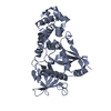
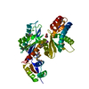
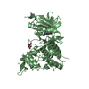
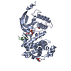
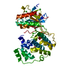
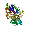
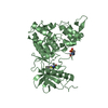
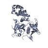
Entry Database : PDB / ID : 6cb0Title Crystal Structure of the FAK FERM domain Focal adhesion kinase 1 Keywords / / Function / homology Function Domain/homology Component
/ / / / / / / / / / / / / / / / / / / / / / / / / / / / / / / / / / / / / / / / / / / / / / / / / / / / / / / / / / / / / / / / / / / / / / / / / / / / / / / / / / / / / / / / / / / / / / / / / / / / / / / / / / / / / Biological species Gallus gallus (chicken)Method / / / Resolution : 1.97 Å Authors Dementiev, A. / Marlowe, T. Journal : BMC Mol Cell Biol / Year : 2019Title : High resolution crystal structure of the FAK FERM domain reveals new insights on the Druggability of tyrosine 397 and the Src SH3 binding site.Authors : Marlowe, T. / Dementiev, A. / Figel, S. / Rivera, A. / Flavin, M. / Cance, W. History Deposition Feb 1, 2018 Deposition site / Processing site Revision 1.0 Feb 6, 2019 Provider / Type Revision 1.1 Jun 5, 2019 Group / Database references / Category / citation_authorItem _citation.journal_abbrev / _citation.journal_id_CSD ... _citation.journal_abbrev / _citation.journal_id_CSD / _citation.journal_id_ISSN / _citation.journal_volume / _citation.page_first / _citation.page_last / _citation.pdbx_database_id_DOI / _citation.pdbx_database_id_PubMed / _citation.title / _citation.year Revision 1.2 Oct 4, 2023 Group / Database references / Refinement descriptionCategory chem_comp_atom / chem_comp_bond ... chem_comp_atom / chem_comp_bond / citation / database_2 / pdbx_initial_refinement_model Item / _database_2.pdbx_DOI / _database_2.pdbx_database_accession
Show all Show less
 Movie
Movie Controller


 Open data
 Basic information
 Components
 Keywords
 Function and homology information
 X-RAY DIFFRACTION /
  Authors
 Citation
 Structure visualization
 Downloads & links
 Other downloads
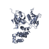


 PDBj
PDBj
 Assembly


 Mass: 18.015 Da / Num. of mol.: 399 / Source method: isolated from a natural source / Formula: H2O
 Sample preparation
 / Beamline: 21-ID-D / Wavelength: 0.9787 Å
 Processing