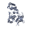
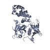
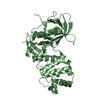
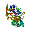
Entry Database : PDB / ID : 4cyeTitle Crystal structure of avian FAK FERM domain FAK31-405 at 3.2A FOCAL ADHESION KINASE 1 Keywords / / / Function / homology Function Domain/homology Component
/ / / / / / / / / / / / / / / / / / / / / / / / / / / / / / / / / / / / / / / / / / / / / / / / / / / / / / / / / / / / / / / / / / / / / / / / / / / / / / / / / / / / / / / / / / / / / / / / / / / / / / / / / / / / / Biological species GALLUS GALLUS (chicken)Method / / / Resolution : 3.2 Å Authors Goni, G.M. / Epifano, C. / Boskovic, J. / Camacho-Artacho, M. / Zhou, J. / Martin, M.T. / Eck, M.J. / Kremer, L. / Graeter, F. / Gervasio, F.L. ...Goni, G.M. / Epifano, C. / Boskovic, J. / Camacho-Artacho, M. / Zhou, J. / Martin, M.T. / Eck, M.J. / Kremer, L. / Graeter, F. / Gervasio, F.L. / Perez-Moreno, M. / Lietha, D. Journal : Proc.Natl.Acad.Sci.USA / Year : 2014Title : Phosphatidylinositol 4,5-Bisphosphate Triggers Activation of Focal Adhesion Kinase by Inducing Clustering and Conformational Changes.Authors : Goni, G.M. / Epifano, C. / Boskovic, J. / Camacho-Artacho, M. / Zhou, J. / Bronowska, A. / Martin, M.T. / Eck, M.J. / Kremer, L. / Grater, F. / Gervasio, F.L. / Perez-Moreno, M. / Lietha, D. History Deposition Apr 10, 2014 Deposition site / Processing site Revision 1.0 Apr 23, 2014 Provider / Type Revision 1.1 Jul 30, 2014 Group Revision 1.2 Aug 20, 2014 Group Revision 1.3 Dec 20, 2023 Group Data collection / Database references ... Data collection / Database references / Other / Refinement description Category chem_comp_atom / chem_comp_bond ... chem_comp_atom / chem_comp_bond / database_2 / pdbx_database_status / pdbx_initial_refinement_model Item / _database_2.pdbx_database_accession / _pdbx_database_status.status_code_sf
Show all Show less
 Movie
Movie Controller


 Open data
 Basic information
 Components
 Keywords
 Function and homology information
 X-RAY DIFFRACTION /
  Authors
 Citation
 Structure visualization
 Downloads & links
 Other downloads


 PDBj
PDBj
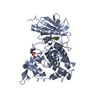
 Assembly
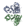

 Mass: 18.015 Da / Num. of mol.: 46 / Source method: isolated from a natural source / Formula: H2O
 Sample preparation
 / Beamline: X06SA / Wavelength: 0.99988
  Processing