Resolution: 2→2.07 Å / Mean I/σ(I) obs: 5.5 / Rsym value: 0.196 / % possible all: 98.7
-
Processing
Software
Name
Version
Classification
CNS
1.1
refinement
DENZO
datareduction
SCALEPACK
datascaling
MLPHARE
phasing
Refinement
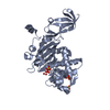
Method to determine structure: SIRAS, MOLECULAR REPLACEMENT Starting model: Arp/Warp autobuilt model using phases from two Ta6Br12 clusters and a poor molecular replacement solution. MR was carried out using a model derived from PDB entry 1HD8, which is a ...Starting model: Arp/Warp autobuilt model using phases from two Ta6Br12 clusters and a poor molecular replacement solution. MR was carried out using a model derived from PDB entry 1HD8, which is a mutant of PBP5, sharing ~26% identity with PBP4 over a 245 residue range. Phase combination with MR solution was necessary, as Ta6Br12 clusters occupied positions on the symmetry planes resulting in centrosymmetric phases and hence uninterpretable maps.
Resolution: 2→23.68 Å / Rfactor Rfree error: 0.003 / Data cutoff high absF: 425919.56 / Data cutoff low absF: 0 / Isotropic thermal model: RESTRAINED / Cross valid method: THROUGHOUT / σ(F): 0 / Stereochemistry target values: Engh & Huber Details: Unidentified electron density is observed near the active site.
In the structure databanks used in Yorodumi, some data are registered as the other names, "COVID-19 virus" and "2019-nCoV". Here are the details of the virus and the list of structure data.
Jan 31, 2019. EMDB accession codes are about to change! (news from PDBe EMDB page)
EMDB accession codes are about to change! (news from PDBe EMDB page)
The allocation of 4 digits for EMDB accession codes will soon come to an end. Whilst these codes will remain in use, new EMDB accession codes will include an additional digit and will expand incrementally as the available range of codes is exhausted. The current 4-digit format prefixed with “EMD-” (i.e. EMD-XXXX) will advance to a 5-digit format (i.e. EMD-XXXXX), and so on. It is currently estimated that the 4-digit codes will be depleted around Spring 2019, at which point the 5-digit format will come into force.
The EM Navigator/Yorodumi systems omit the EMD- prefix.
Related info.:Q: What is EMD? / ID/Accession-code notation in Yorodumi/EM Navigator
Yorodumi is a browser for structure data from EMDB, PDB, SASBDB, etc.
This page is also the successor to EM Navigator detail page, and also detail information page/front-end page for Omokage search.
The word "yorodu" (or yorozu) is an old Japanese word meaning "ten thousand". "mi" (miru) is to see.
Related info.:EMDB / PDB / SASBDB / Comparison of 3 databanks / Yorodumi Search / Aug 31, 2016. New EM Navigator & Yorodumi / Yorodumi Papers / Jmol/JSmol / Function and homology information / Changes in new EM Navigator and Yorodumi
 Movie
Movie Controller

 Yorodumi
 Open data
 Basic information
 Components
 Keywords
 Function and homology information
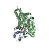
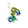
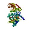
 Staphylococcus aureus (bacteria)
 X-RAY DIFFRACTION /
  Authors
 Citation
 Structure visualization
 Downloads & links
 Other downloads
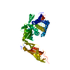


 PDBj
PDBj


 Assembly


 Mass: 96.063 Da / Num. of mol.: 5 / Source method: obtained synthetically / Formula: SO4
 Mass: 18.015 Da / Num. of mol.: 781 / Source method: isolated from a natural source / Formula: H2O
 Sample preparation
 Processing