 Movie
Movie Controller

[English] 日本語
 Yorodumi
- PDB-3tzy: Crystal structure of a fragment containing the acyltransferase do... -
+ Open data

- Basic information
| Entry | Database: PDB / ID: 3tzy | ||||||
|---|---|---|---|---|---|---|---|
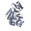
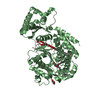
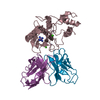
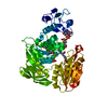
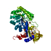
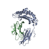
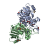
| Title | Crystal structure of a fragment containing the acyltransferase domain of Pks13 from Mycobacterium tuberculosis in the palmitoylated form at 2.2 A | ||||||
 Components Components |
| ||||||
 Keywords Keywords | TRANSFERASE / Acyltransferase / Long fatty acid chain transferase / Acyl carrier protein | ||||||
| Function / homology |  Function and homology information Function and homology informationpolyketide synthase complex / fatty acid elongation, saturated fatty acid / mycolate cell wall layer assembly / mycolic acid biosynthetic process / DIM/DIP cell wall layer assembly / acyltransferase activity, transferring groups other than amino-acyl groups / fatty acid synthase activity / phosphopantetheine binding / 3-oxoacyl-[acyl-carrier-protein] synthase activity / peptidoglycan-based cell wall ...polyketide synthase complex / fatty acid elongation, saturated fatty acid / mycolate cell wall layer assembly / mycolic acid biosynthetic process / DIM/DIP cell wall layer assembly / acyltransferase activity, transferring groups other than amino-acyl groups / fatty acid synthase activity / phosphopantetheine binding / 3-oxoacyl-[acyl-carrier-protein] synthase activity / peptidoglycan-based cell wall / protein homooligomerization / plasma membrane / cytosol Similarity search - Function | ||||||
| Biological species |   Mycobacterium tuberculosis (bacteria) Mycobacterium tuberculosis (bacteria) | ||||||
| Method |  X-RAY DIFFRACTION / SYNCHROTRON / MOLECULAR REPLACEMENT / Resolution: 2.2 Å X-RAY DIFFRACTION / SYNCHROTRON / MOLECULAR REPLACEMENT / Resolution: 2.2 Å | ||||||
 Authors Authors | Bergeret, F. / Pedelacq, J.D. / Mourey, L. / Bon, C. | ||||||
 Citation Citation | Journal: J.Biol.Chem. / Year: 2012 Title: Biochemical and structural study of the atypical acyltransferase domain from the mycobacterial polyketide synthase pks13 Authors: Bergeret, F. / Gavalda, S. / Chalut, C. / Malaga, W. / Quemard, A. / Pedelacq, J.D. / Daffe, M. / Guilhot, C. / Mourey, L. / Bon, C. | ||||||
| History |
|
- Structure visualization
| Structure viewer | Molecule: MolmilJmol/JSmol |
|---|
- Downloads & links
-Download
| PDBx/mmCIF format | 3tzy.cif.gz | 196.9 KB | Display | PDBx/mmCIF format |
|---|---|---|---|---|
| PDB format | pdb3tzy.ent.gz | 154.3 KB | Display | PDB format |
| PDBx/mmJSON format | 3tzy.json.gz | Tree view | PDBx/mmJSON format | |
| Others |  Other downloads Other downloads |
-Validation report
| Arichive directory | https://data.pdbj.org/pub/pdb/validation_reports/tz/3tzyftp://data.pdbj.org/pub/pdb/validation_reports/tz/3tzy | HTTPS FTP |
|---|
-Related structure data
| Related structure data |  3tzwC  3tzxC 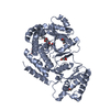 3tzzC 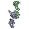 2hg4S C: citing same article ( S: Starting model for refinement |
|---|---|
| Similar structure data |


-Links
 PDBj
PDBj


- Assembly
| Deposited unit | 
| ||||||||
|---|---|---|---|---|---|---|---|---|---|
| 1 | 
| ||||||||
| 2 | 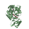
| ||||||||
| Unit cell |
| ||||||||
| Details | THE HETERODIMER FORMED BETWEEN THE UNKNOWN PEPTIDE AND THE ACYLTRANSFERASE DOMAIN OF PKS13 HAS NO KNOWN FUNCTIONAL RELEVANCE FOR THE TIME BEING |
-Components
-Protein / Protein/peptide , 2 types, 4 molecules ABCD
| #1: Protein | Mass: 53086.805 Da / Num. of mol.: 2 / Fragment: Acyltransferase domain, UNP residues 576-1062 Source method: isolated from a genetically manipulated source Source: (gene. exp.) Mycobacterium tuberculosis (bacteria) / Strain: H37Rv / Gene: Rv3800c / Plasmid: pWM71, pET28aII / Production host: References: UniProt: O53579, Transferases; Acyltransferases; Transferring groups other than aminoacyl groups #2: Protein/peptide | Mass: 1355.495 Da / Num. of mol.: 2 / Source method: isolated from a natural source Details: The author presume that this peptide comes from the Escherichia coli strain that was used to produce the recombinant protein. Source: (natural) |
|---|
-Non-polymers , 4 types, 245 molecules 


| #3: Chemical |  Mass: 256.424 Da / Num. of mol.: 2 / Source method: obtained synthetically / Formula: C16H32O2 Mass: 256.424 Da / Num. of mol.: 2 / Source method: obtained synthetically / Formula: C16H32O2#4: Chemical | ChemComp-GOL /  Mass: 92.094 Da / Num. of mol.: 7 / Source method: obtained synthetically / Formula: C3H8O3 Mass: 92.094 Da / Num. of mol.: 7 / Source method: obtained synthetically / Formula: C3H8O3#5: Chemical |  Mass: 96.063 Da / Num. of mol.: 2 / Source method: obtained synthetically / Formula: SO4 Mass: 96.063 Da / Num. of mol.: 2 / Source method: obtained synthetically / Formula: SO4#6: Water | ChemComp-HOH / | Mass: 18.015 Da / Num. of mol.: 234 / Source method: isolated from a natural source / Formula: H2O |
|---|
-Details
| Has protein modification | Y |
|---|
-Experimental details
-Experiment
| Experiment | Method: X-RAY DIFFRACTION / Number of used crystals: 1 |
|---|
- Sample preparation
| Crystal | Density Matthews: 3.34 Å3/Da / Density % sol: 63.19 % |
|---|---|
| Crystal grow | Temperature: 285 K / Method: vapor diffusion, hanging drop / pH: 7.5 Details: 0.1M HEPES, 1.7M ammonium sulfate, 15% glycerol, 1.7% PEG-400, pH 7.5, VAPOR DIFFUSION, HANGING DROP, temperature 285K |
-Data collection
| Diffraction | Mean temperature: 100 K |
|---|---|
| Diffraction source | Source: SYNCHROTRON / Site: ESRF  / Beamline: ID14-4 / Wavelength: 0.9395 Å / Beamline: ID14-4 / Wavelength: 0.9395 Å |
| Detector | Type: ADSC QUANTUM 315r / Detector: CCD / Date: Mar 7, 2009 |
| Radiation | Protocol: SINGLE WAVELENGTH / Monochromatic (M) / Laue (L): M / Scattering type: x-ray |
| Radiation wavelength | Wavelength: 0.9395 Å / Relative weight: 1 |
| Reflection | Resolution: 2.2→52.9 Å / Num. obs: 69116 / % possible obs: 91.5 % / Redundancy: 7 % / Biso Wilson estimate: 42.2 Å2 / Rsym value: 0.07 |
| Reflection shell | Resolution: 2.2→2.3 Å / Mean I/σ(I) obs: 2.4 / Rsym value: 0.316 / % possible all: 89.5 |
- Processing
| Software |
| ||||||||||||||||||||||||||||||||||||||||||||||||||||||||||||||||||||||||||||||||||||||||||||||||||||||||||||||||||||||||||||||||||||||||||||||||||||||||||||||||||||||||||
|---|---|---|---|---|---|---|---|---|---|---|---|---|---|---|---|---|---|---|---|---|---|---|---|---|---|---|---|---|---|---|---|---|---|---|---|---|---|---|---|---|---|---|---|---|---|---|---|---|---|---|---|---|---|---|---|---|---|---|---|---|---|---|---|---|---|---|---|---|---|---|---|---|---|---|---|---|---|---|---|---|---|---|---|---|---|---|---|---|---|---|---|---|---|---|---|---|---|---|---|---|---|---|---|---|---|---|---|---|---|---|---|---|---|---|---|---|---|---|---|---|---|---|---|---|---|---|---|---|---|---|---|---|---|---|---|---|---|---|---|---|---|---|---|---|---|---|---|---|---|---|---|---|---|---|---|---|---|---|---|---|---|---|---|---|---|---|---|---|---|---|---|
| Refinement | Method to determine structure: MOLECULAR REPLACEMENT Starting model: 2HG4 Resolution: 2.2→50 Å / Cor.coef. Fo:Fc: 0.943 / Cor.coef. Fo:Fc free: 0.921 / SU B: 6.023 / SU ML: 0.151 / Cross valid method: THROUGHOUT / ESU R: 0.234 / ESU R Free: 0.206 / Stereochemistry target values: MAXIMUM LIKELIHOOD
| ||||||||||||||||||||||||||||||||||||||||||||||||||||||||||||||||||||||||||||||||||||||||||||||||||||||||||||||||||||||||||||||||||||||||||||||||||||||||||||||||||||||||||
| Solvent computation | Ion probe radii: 0.8 Å / Shrinkage radii: 0.8 Å / VDW probe radii: 1.2 Å / Solvent model: MASK | ||||||||||||||||||||||||||||||||||||||||||||||||||||||||||||||||||||||||||||||||||||||||||||||||||||||||||||||||||||||||||||||||||||||||||||||||||||||||||||||||||||||||||
| Displacement parameters | Biso mean: 44.418 Å2
| ||||||||||||||||||||||||||||||||||||||||||||||||||||||||||||||||||||||||||||||||||||||||||||||||||||||||||||||||||||||||||||||||||||||||||||||||||||||||||||||||||||||||||
| Refinement step | Cycle: LAST / Resolution: 2.2→50 Å
| ||||||||||||||||||||||||||||||||||||||||||||||||||||||||||||||||||||||||||||||||||||||||||||||||||||||||||||||||||||||||||||||||||||||||||||||||||||||||||||||||||||||||||
| Refine LS restraints |
| ||||||||||||||||||||||||||||||||||||||||||||||||||||||||||||||||||||||||||||||||||||||||||||||||||||||||||||||||||||||||||||||||||||||||||||||||||||||||||||||||||||||||||
| LS refinement shell | Resolution: 2.2→2.257 Å / Total num. of bins used: 20
|
